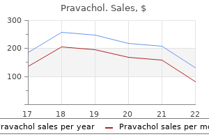
Pravachol

Pravachol
Pravachol dosages: 20 mg, 10 mg
Pravachol packs: 30 pills, 60 pills, 90 pills, 120 pills, 180 pills, 270 pills

In these injuries cholesterol levels uk 5.3 discount pravachol 10mg amex, restore results from exercise of the pluripotential progenitor cells located within the perichondrium cholesterol test at home pravachol 20 mg purchase on-line. At the molecular degree cholesterol in eggs is good pravachol 10mg buy discount, cartilage restore is a tentative stability between deposition of kind I collagen within the type of scar tissue and restore by expression of the cartilage-specific collagens. However, in adults, new blood vessels generally develop at the web site of the therapeutic wound that stimulate the growth of bone quite than actual cartilage restore. The limited ability of cartilage to restore itself could cause vital problems in cardiothoracic surgical procedure, such as coronary artery bypass graft surgical procedure, when costal cartilage should be cut to enter the chest cavity. Hyaline cartilage is prone to calcification, a course of in which calcium phosphate crystals turn into embedded in the cartilage matrix. B In most of those conditions, given adequate time, cartilage that calcifies is replaced by bone. Chondrocytes normally derive all of their nutrients and dispose of wastes by diffusion of supplies by way of the matrix. When the matrix turns into closely calcified, diffusion is impeded and the chondrocytes swell and die. The final consequence of this event is removal of the calcified matrix and its alternative by bone. A number of investigators believe the method of cartilage elimination entails a selected cell sort designated as a chondroclast. This cell is described as resembling an osteoclast in both morphology and lytic operate. Chondroclasts have additionally been observed on the deep floor of resorbed articular cartilage in numerous joint illnesses. For instance, these multinucleated cells have been recognized on both calcified and noncalcified articular cartilage erosions in rheumatoid arthritis. Recent immunocytochemical research on chondroclasts obtained from pathological joint specimens revealed that chondroclasts express the osteoclast-type phenotype. It is in all probability going that chondroclasts are mature osteoclasts, which are capable to resorb cartilage and are discovered wherever cartilage is being eliminated. The darker, somewhat basophilic areas on the left side of the micrograph represent normal cartilage matrix (C). The lighter and more eosinophilic areas characterize bone tissue (B) that has changed the unique cartilage matrix. A giant marrow cavity has formed throughout the cartilage construction and is seen within the center of the micrograph. Cartilage is an avascular construction; therefore, the composition of the extracellular matrix is crucial for diffusion of substances between chondrocytes and blood vessels within the surrounding connective tissue. There are three main forms of cartilage: hyaline cartilage, elastic cartilage, and fibrocartilage. Chondrocytes are distributed both singularly or in clusters known as isogenous teams. Extracellular matrix surrounding particular person chondrocytes (capsular matrix) or the isogenous group (territorial matrix) varies in collagen content and staining properties. The interterritorial matrix surrounds the territorial cartilage is produced by chondrocytes and seems glassy. Hyaluronan molecules work together with numerous aggrecan molecules to form large proteoglycan matrix and occupies the space between isogenous teams. Hyaline cartilage is a key tissue within the growth of the fetal skeleton (endochondral ossification) and in most rising bones (epiphyseal progress plates). Fibrocartilage is typically current in intervertebral discs, the pubic symphysis, insertion of tendons, and structures within certain joints. In addition, floor substance accommodates larger quantities of versican than aggrecan molecules. Repair largely (forms new cartilage at the surface of an present cartilage) and interstitial growth (forms new cartilage by mitotic division of chondrocytes within an current cartilage mass). In the getting older course of, hyaline cartilage is prone to calcification and is changed by bone. All collagen molecules work together with each other in a three-dimensional felt-like arrangement. The matrix is highly hydrated-more than 60% of its web weight consists of water, most of which is bound to proteoglycan aggregates (aggrecan monomers sure to an extended hyaluronan molecule). In addition, hyaline cartilage constitutes much of the fetal skeleton and performs an essential position within the progress of most bones. At most websites within the physique, except for synovial joint surfaces, hyaline cartilage is surrounded by dense irregular connective tissue known as the perichondrium. Hyaline cartilage displays both appositional progress, the addition of recent cartilage at its floor by chondroblasts, and interstitial growth, the division and differentiation of chondrocytes inside its extracellular matrix. The newly divided cells produce new cartilage matrix, thus increasing the amount of the cartilage from inside. Therefore, the overall growth of cartilage results from the interstitial secretion of latest matrix by chondrocytes and by the appositional secretion of matrix by newly differentiated chondroblasts. This micrograph reveals hyaline cartilage from the trachea as seen in a routinely prepared specimen. The cartilage appears as an avascular expanse of matrix material and a population of cells known as chondrocytes (Ch). The chondrocytes produce the matrix; the house every chondrocyte occupies is called a lacuna (L). The perichondrium serves as a supply of new chondrocytes throughout appositional growth of the cartilage. Often, the perichondrium reveals two distinctive layers: an outer, more fibrous layer and an inner, extra cellular layer. The inner, more cellular layer, containing chondroblasts and chondroprogenitor cells, supplies for external development. The matrix additionally incorporates, amongst other elements, sulfated glycosaminoglycans that exhibit basophilia with hematoxylin or other fundamental dyes. Also, the matrix material immediately surrounding a lacuna tends to stain extra intensely with primary dyes. Not uncommonly, the matrix might seem to stain more intensely in localized areas (asterisks) that look much like the capsule matrix. This outcomes from inclusion of a capsule inside the thickness of the part however not the lacuna it surrounds. Frequently, two or more chondrocytes are located extraordinarily near one another, separated by solely a skinny partition of matrix. The proliferation of recent chondrocytes by this means with the resultant addition of matrix results in interstitial growth of the cartilage. Also, notice the very distinct and deeply stained capsules (arrows) surrounding the chondrocytes. The capsule represents the site the place the sulfated glycosaminoglycans are most concentrated. In contrast to the basophilia of the cartilage matrix, the perichondrium (P) is stained with eosin. The frivolously stained region between the perichondrium and the deeply stained matrix is matrix that has not but matured. The hyaline cartilage on this micrograph is from a specimen obtained shortly after death and kept cool during fixation. The procedure reduces the lack of its negatively charged sulfate groups; thus, the matrix is stained extra heavily with Hyaline cartilage, trachea, human, H&E 850. This larger magnification micrograph reveals the area inside the rectangle within the lower left figure. The chondrocytes (Ch) within the upper a part of the micrograph characterize an isogenous group and are producing matrix materials for interstitial development. The lightly stained basophilic area reveals immature chondrocytes (arrows) within the perichondrium (P). These cells are formative chondrocytes that are simply beginning to , or will shortly, produce matrix material. In distinction, the nuclei near the underside fringe of the micrograph are fibroblast nuclei (Fib); they belong to the outer layer of the perichondrium. Note how attenuated their nuclei are compared with the formative chondroblast nuclei of the inside perichondrial layer. This cartilage is replaced by bone tissue besides the place one bone contacts another, as in a movable joint. In these places, cartilage persists and covers the top of each bone as articular cartilage, providing a easy, well-lubricated surface towards which the end of one bone moves on the opposite within the joint.
Diseases
This laser-scanning confocal image of protoplasmic astrocyte in the gray matter of the dentate gyrus was visualized by intracellular labeling method what kind of cholesterol in eggs trusted 20 mg pravachol. In flippantly mounted tissue slices cholesterol medication body odor pravachol 10 mg proven, selected astrocytes have been impaled and iontophoretically injected with fluorescent dye (Alexa Fluor 568) using pulses of adverse present xylitol cholesterol generic pravachol 10 mg without a prescription. Examination of the connection between astrocyte morphology and laminar boundaries in the molecular layer of adult dentate gyrus. Photomicrograph of the white matter of the mind, showing the in depth radiating cytoplasmic processes for which astrocytes are named. They assist keep the tight junctions of the capillaries that type the blood�brain barrier (see web page 388). In addition, astrocytes present a overlaying for the "naked areas" of myelinated axons- for example, on the nodes of Ranvier and at synapses. They could confine neurotransmitters to the synaptic cleft and take away extra neurotransmitters by pinocytosis. This diagram exhibits the 4 types of glial cells-astrocytes, oligodendrocytes, microglial cells, and ependymal cells-interacting with several structures and cells found in the brain tissue. Note that the astrocytes and their processes interact with the blood vessels in addition to with axons and dendrites. Astrocytes also ship their processes towards the mind surface, the place they contact the basement membrane of the pia mater, forming the glia limitans. The astrocyte plasma membrane accommodates an abundance of K pumps and K channels that mediate the transfer K ions from areas of high to low focus. Accumulation of enormous amounts of intracellular K in astrocytes decreases native extracellular K gradients. The astrocyte membrane turns into depolarized, and the cost is dissipated over a large space by the in depth community of astrocyte processes. Instead, every tongue-like course of appears to spiral across the axon, always staying in proximity to it, till the myelin sheath is formed. Oligodendrocytes seem in specifically stained mild microscopic preparations as small cells with comparatively few processes compared with astrocytes. Each oligodendrocyte offers off several tongue-like processes that discover their approach to the axons, the place each process wraps itself round a portion of an axon, forming an internodal segment of myelin. The nucleus-containing region of the oligodendrocyte may be at a long way from the axons it myelinates. Thus, where myelin sheaths of adjacent axons touch, they could share an intraperiod line. Cytoplasmic processes from the oligodendrocyte cell physique form flattened cytoplasmic sheaths that wrap around each of the axons. The relationship of cytoplasm and myelin is essentially the same as that of Schwann cells. Microglial cells are considered a half of the mononuclear phagocytotic system (see Folder 6. Recent evidence suggests that microglia play a critical role in defense towards invading microorganisms and neoplastic cells. They take away bacteria, injured cells, and the debris of cells that undergo apoptosis. They also mediate neuroimmune reactions, corresponding to those occurring in chronic ache situations. Photomicrograph of microglial cells (arrows) showing their attribute elongated nuclei. In this condition, the microglial cells are present in giant numbers and are readily seen in a routine H&E preparation. The spikes will be the equivalent of the ruffled border seen on different phagocytotic cells. Ependymal cells form the epithelial-like lining of the ventricles of the brain and spinal canal. The cell physique of tanycytes offers rise to a long course of that tasks into the mind parenchyma. Tanycytes are delicate to glucose concentration; due to this fact, they may be involved in detecting and responding to modifications in vitality stability in addition to in monitoring different circulating metabolites in the cerebrospinal fluid. Photomicrograph of the central area of the spinal wire stained with toluidine blue. At higher magnification, ependymal cells, which line the central canal, can be seen to encompass a single layer of columnar cells. Transmission electron micrograph exhibiting a portion of the apical region of two columnar ependymal cells. The modified ependymal cells and associated capillaries are referred to as the choroid plexus. Impulse Conduction An motion potential is an electrochemical course of triggered by impulses carried to the axon hillock after different impulses are acquired on the dendrites or the cell body itself. In response to a stimulus, voltage-gated Na channels in the preliminary segment of the axon membrane open, causing an influx of Na into the axoplasm. This inflow of Na briefly reverses (depolarizes) the negative membrane potential of the resting membrane (70 mV) to optimistic (30 mV). After depolarization, the voltage-gated Na channels shut and voltagegated K channels open. Depolarization of 1 part of the membrane sends electrical current to neighboring portions of unstimulated membrane, which is still positively charged. After a really temporary (refractory) period, the neuron can repeat the method of generating an action potential as soon as again. Physiologists describe the nerve impulse as "leaping" from node to node alongside the myelinated axon. However, the voltage reversal can solely happen on the nodes of Ranvier, where the axolemma lacks a myelin sheath. Because of this, the voltage reversal (and, thus, the impulse) jumps as present flows from one node of Ranvier to the subsequent. The speed of saltatory conduction is said not only to the thickness of the myelin but also to the diameter of the axon. In unmyelinated axons, Na and K channels are distributed uniformly alongside the size of the fiber. The nerve impulse is carried out more slowly and strikes as a continuous wave of voltage reversal alongside the axon. However, in the adult mammalian mind, a really small variety of cells left from improvement referred to as neural stem cells retain the flexibility to divide. These cells migrate into sites of harm and differentiate into totally functional nerve cells. They seem to share a developmental lineage with motor neurons migrating from their site of origin to creating axonal projections (tracts) in the white matter of the brain or spinal twine. The precursors then proliferate in response to the native expression of mitogenic signals. The matching of oligodendrocytes to axons is completed by way of a mix of native regulation of cell proliferation, differentiation, and apoptosis. During the embryonic and early postnatal stages, immature astrocytes migrate into the cortex, where they differentiate and become mature astrocytes. Ependymal cells are derived from the proliferation of neuroepithelial cells that immediately surround the canal of the growing neural tube. As the one glial cells of mesenchymal origin, microglia possess the vimentin class of intermediate filaments, which can be useful in identifying these cells with immunocytochemical strategies. Neurons, oligodendrocytes, astrocytes, and ependymal cells are derived from cells of the neural tube. Schwann cells additionally arise from migrating neural crest cells that become associated with the axons of early embryonic nerves. Axon-derived neuregulin 1 (Nrg-1) sustains the Schwann cell precursors that endure differentiation and divide alongside the rising nerve processes. Immature Schwann cells that affiliate with large-diameter axons mature into myelinating Schwann cells, whereas those who associate with small-diameter axons mature into nonmyelinating cells. The cell our bodies of motor neurons that innervate skeletal muscle (somatic efferents) are situated in the brain, mind stem, and spinal cord. It can connote the axon with all of its coverings (myelin and Schwann cell), as used above, or it can connote the axon alone.
This explains why wounds fail to heal and bone formation is impaired in scurvy (vitamin C deficiency) cholesterol test perth pravachol 20mg quality. O-linked sugar teams are added to some hydroxylysine residues (glycosylation) cholesterol normal lab values proven 20 mg pravachol, and N-linked sugars are added to the two terminal positions cholesterol chart printable cheap pravachol 10 mg fast delivery. The globular construction is formed on the carboxyterminus, which is stabilized by disulfide bonds. Formation of this construction ensures the right alignment of the three chains during the formation of the triple helix. A triple helix (beginning from the carboxy-terminus) is shaped by three chains, except at the terminals the place the polypeptide chains remain uncoiled. Intrachain and interchain hydrogen and disulfide bonds kind that affect the form of the molecule. The triple-helix molecule is stabilized by the binding of the chaperone protein hsp47, which additionally prevents the untimely aggregation of the trimers within the cell. The folded procollagen molecules cross to the Golgi equipment and begin to affiliate into small bundles. This bundling is achieved by the lateral associations between uncoiled terminals of the procollagen molecules. Free and small aggregates of procollagen molecules are packaged into secretory vesicles and transported to the cell surface. The aggregated collagen molecules then align together to type the ultimate collagen fibrils in a course of generally recognized as fibrillogenesis. The cell controls the orderly array of the newly formed fibrils by directing the secretory vesicles to a localized surface site for discharge. Within the cove, the collagen molecules align in rows and self-assemble longitudinally in a head-to-tail trend. Scissors within the high illustration show the place C and N terminals are cleaved by carboxy- and aminopeptidase, respectively, from the procollagen molecule to kind the collagen molecule. On the C terminus of the molecule, the sugar subunit is GlcNac (N-acetylglucosamine) connected to mannose (Man)n. Globular N-terminal propeptide is smaller and has quick triple-helical and nontriple-helical domains, whereas C-terminal propeptide is larger with a single nontriple-helical area. Collagen biogenesis ends in the formation of extremely organized polymers known as fibrils. The fibrils additional affiliate with one another to form larger collagen fibers, which on a per weight basis have the tensile power comparable to that of metal. For instance, collagen type I fiber of 1 mm in diameter can face up to a load of 10 to forty kg before it breaks. They control the thickness of kind I fibrils by limiting the deposition of collagen molecules after the fibril has reached the specified diameter. Collagen molecules are synthesized by numerous types of connective tissue and epithelial cells. In addition, the collagen molecules of basement membrane (see web page 137) are produced by epithelial cells. The synthesis of collagen is regulated by complicated interactions among growth components, hormones, and cytokines. Initial fragmentation of insoluble collagen molecules occurs through mechanical wear, the motion of free radicals, or proteinase cleavage. The resulting collagen fragments are then phagocytosed by cells and degraded by their lysosomal enzymes. Most collagenopathies are attributed to mutations in genes encoding the chains in the varied collagens. Mutation of collagens produces a extensive variety of genetic problems that vary from gentle to lethal, depending on the mutation of the collagen gene and its subsequent effect on the molecular construction of the collagen and its function in the physique. In the future, gene therapy may probably be used both to management deposition of faulty collagen or to reverse the disease course of attributable to the mutated genes. Fibroblasts are additionally capable of phagocytosing and degrading collagen fibrils throughout the lysosomes of the cell. Reticular Fibers Reticular fibers provide a supporting framework for the mobile constituents of assorted tissues and organs. It surrounds the fiber with its cytoplasm, thus isolating the fiber from other tissue parts. Important exceptions to this common rule include the endoneurium of peripheral nerves, the place Schwann cells secrete reticular fibers; tunica media of blood vessels; and muscularis of the alimentary canal, the place easy muscle cells secrete reticular and other collagen fibers. Connective Tissue Elastic fibers are usually thinner than collagen fibers and are organized in a branching sample to kind a threedimensional network. The fibers are interwoven with collagen fibers to limit the distensibility of the tissue and prevent tearing from excessive stretching (Plate 6, page 192). Because elastic fibers become somewhat refractile with sure fixatives, they might be distinguished from collagen fibers in specimens stained with H&E when they show this attribute. Photomicrograph of a lymph node silver preparation displaying the connective tissue capsule on the high and a trabecula extending from it on the left. When visualized in the gentle microscope with particular methods, reticular fibers have a thread-like look. They are also revealed with special silver-staining procedures such because the Gomori and Wilder strategies. Reticular fibers are named for his or her association in a meshlike sample or network. E C In free connective tissue, networks of reticular fibers are discovered on the boundary of connective tissue and epithelium, as nicely as surrounding adipocytes, small blood vessels, nerves, and muscle cells. As embryonic growth or wound healing progresses, reticular fibers are progressively replaced by the stronger sort I collagen fibers. Reticular fibers additionally perform as a supporting stroma in hemopoietic and lymphatic tissues (but not within the thymus). The mesentery may be very thin, and the microscope may be centered via the complete thickness of the tissue. The elastic property of the elastin molecule is related to its unusual polypeptide spine, which causes random coiling. Elastic fibers are produced by lots of the identical cells that produce collagen and reticular fibers, notably fibroblasts and easy muscle cells. In distinction to collagen fibers, nevertheless, elastic fibers are composed of two structural elements: a central core of elastin and a surrounding network of fibrillin microfibrils. The random distribution of glycines makes the elastin molecule hydrophobic and allows for random coiling of its fibers. This permits elastic fibers to "slide" over one another or to be stretched and then recoil to their original state. Elastin additionally incorporates desmosine and isodesmosine, two giant amino acids distinctive to elastin, which are liable for the covalent bonding of elastin molecules to one another. Elastin varieties fibers of variable thicknesses, or lamellar � layers (as in elastic arteries). The elastin gene consists of 28 kilobases however lower than 10% of the kilobases carry the sequence that encodes elastin. Fibrillin-1 (350 kDa) is a glycoprotein that varieties fine microfibrils measuring 10 to 12 nm in diameter. During the early levels of elastogenesis, fibrillin microfibrils are used as substrates for the meeting of elastic fibers. The microfibrils are fashioned first; elastin material is then deposited on the surface of the microfibrils. Emilin-1 (elastin microfibril interface-located protein, 106 kDa) is one other glycoprotein found at the elastin� fibrillin microfibril interface that most likely regulates the deposition of elastin in the course of the formation of fibers. Both elastin-associated fibrillin microfibrils and emilin-1 play a significant function in regulating elastogenesis. Immunofluorescence of a pores and skin biopsy specimen from a person with this syndrome shows an absence of elastin-associated fibrillin microfibrils. In addition, mutation in the emilin-1 gene locus exhibits alterations of the fantastic structure of elastic fibers and of cell morphology in the elastic arteries. In mature fibers, the fibrillin microfibrils are situated throughout the elastic fiber and at its periphery. The presence of microfibrils inside the fiber is related to the expansion course of; thus, as the fiber is fashioned and thickens, the microfibrils turn out to be entrapped throughout the newly deposited elastin.
The neutrophil to the best exhibits larger maturity by advantage of its very distinctive lobulation cholesterol medication list pravachol 10 mg order line. The eosinophils seen in these micrographs equally characterize different stages of maturity cholesterol diabetes pravachol 10mg order on-line. The eosinophil on the left is comparatively small and is simply beginning to show lobulation cholesterol lab values chart discount pravachol 20mg line. The cytoplasm is nearly entirely filled with eosinophilic granules that characterize this cell kind. The lighter stained space, devoid of granules, most likely represents the site of the Golgi apparatus (arrow). The eosinophil proven within the center is larger and its nucleus is now distinctively bilobed. The eosinophil on the right is extra mature in that it displays at least three lobes. By going by way of focus, the eosinophil granules usually seem to "light up" as a outcome of their crystalline structure. The cells shown listed beneath are basophils and also characterize different stages of maturation. The basophilic granules are variable in dimension and tend to obscure the morphology of the nucleus. The nucleus of the center basophil seems to be bilobed, but the granules that lie over the nucleus again are inclined to obscure the precise form. The distinction in lymphocyte measurement is attributable mostly to the quantity of cytoplasm current. However, the nucleus also contributes to the scale of the cell but to a lesser degree. Their measurement ranges from approximately 13 to 20 m, with the majority falling in the upper dimension range. Small, azurophilic granules (lysosomes) are additionally characteristic of the cytoplasm and are similar to those seen in neutrophils. In preparations corresponding to this, the lipid content is misplaced during preparation and recognition of the cell is based on a clear or unstained round house. The megakaryocyte is a polyploid cell that displays a large and irregular nuclear profile. However, examples of each stage of development in both cell strains are introduced in the following plates. In distinction, many cells in their late stage of development, particularly within the granulocyte sequence, could be identified with some degree of assuredness at low magnification. This kind of preparation allows for the examination of growing pink and white cells. A pattern of bone marrow is aspirated from a bone and easily positioned on a slide and spread into a skinny monolayer of cells. These are very young erythrocytes that include residual ribosomes of their cytoplasm. Their cytoplasm is basophilic and the nucleus displays a dense chromatin construction and several nucleoli. The cytoplasm shows robust basophilia due to the increasing variety of ribosomes concerned in hemoglobin synthesis. The accumulation of hemoglobin in the cell steadily changes the staining response of the cytoplasm in order that it begins to stain with eosin. The recognizable presence of hemoglobin within the cell by virtue of its staining signifies its transition to the polychromatophilic erythroblast. With time, increasing quantities of hemoglobin are synthesized and concomitantly, lowering numbers of ribosomes are present. The nucleus of the cell is smaller than that of the basophilic erythroblast and the heterochromatin is far coarser. At the top of this stage, the nucleus has become a lot smaller and the cytoplasm more eosinophilic. The next definable stage is the orthochromatophilic erythroblast, additionally called normoblast. The cytoplasm is considerably much less blue leaning more to a pink or eosinophilic coloration. In the subsequent stage, the polychromatophilic erythrocyte, also extra commonly known as a reticulocyte, has lost its nucleus and is prepared to pass into the blood sinusoids of the pink bone marrow. Comparison of this cell to typical mature erythrocytes within the marrow smear reveals a slight distinction in coloration. The greater abundance of cytoplasm is deeply basophilic compared to that of the proerythroblast. The cytoplasm is basophilic however is significantly lighter in colour than that of the basophilic erythroblast. The cytoplasm additionally reveals some eosinophilia, which is indicative of hemoglobin production. Note how rather more dense the chromatin appears in addition to how a lot smaller the nucleus has turn out to be. The cytoplasm is predominantly eosinophilic but nonetheless possesses a degree of basophilia. Their nuclei have turn out to be even smaller Polychromatophilic erythrocyte, bone marrow smear, human, Giemsa, 2,200. Compare the coloration of the polychromatophilic erythrocyte with that of the mature red blood cells. Polychromatophilic erythrocytes can additionally be readily demonstrated with particular stains that cause the remaining ribosomes within the cytoplasm to clump and kind a visible reticular community, therefore, the polychromatophilic erythrocyte is also commonly referred to as a reticulocyte. The earliest recognizable stage is the myeloblast, which is followed consecutively by the promyelocyte, myelocyte, metamyelocyte, band cell, and at last, the mature granulocyte. The cells of the basophil lineage are extremely tough to locate in a marrow smear due to the minimal variety of these cells within the marrow. The myeloblast is characterized by a large euchromatic, spherical nucleus with three to 5 nucleoli. The cytoplasm of the neutrophilic myelocyte is characterised by small, pink-to-red specific granules with some azurophilic granules current. The eosinophilic lineage has a similar-appearing nucleus, however its specific granules are giant. The nuclear-cytoplasmic ratio is additional decreased and the nucleus assumes a kidney shape. The eosinophilic metamyelocyte reveals an elevated number of specific granules compared to the neutrophilic metamyelocyte. The chromatin of the nucleus reveals further condensation and has a horseshoe form. In the neutrophilic band cell, the small, pink-to-red specific granules are the one granule kind present. The eosinophilic band cell shows little or no change relative to the precise granules, however the nucleus reveals a kidney shape. The myeloblast proven right here reveals a deep blue cytoplasm with a lighter region that represents the Golgi area (G). The eosinophilic myelocyte displays a nucleus the identical as that described for the neutrophilic myelocyte. The neutrophilic myelocyte retains the round nucleus, but nucleoli are actually absent. The cytoplasm reveals quite a few characteristic eosinophilic granules which might be present throughout the cytoplasm. The neutrophilic metamyelocyte differs from its precursor by the presence of a kidney- or bean-shaped nucleus. The small, pink-to-red particular granules are now seen within the cytoplasm and few or no azurophilic granules are present. The band or nonsegmented neutrophil reveals a horseshoe-shaped nucleus with abundant small, pink-to-red specific granules within the cytoplasm. In contrast, muscle cells contain a lot of aligned contractile filaments that the cells use for the only purpose of manufacturing mechanical work.
Red Malaga (Grape). Pravachol.
Source: http://www.rxlist.com/script/main/art.asp?articlekey=96481
The upper floor of the cantilever is reflective cholesterol ratio american heart association order pravachol 10mg otc, and a laser beam is directed off the cantilever to a diode cholesterol medication during pregnancy 20 mg pravachol buy with visa. This association acts as an "optical lever" because extraordinarily small deflections of the cantilever are tremendously magnified on the diode cholesterol lowering foods with added plant sterols cheap pravachol 20mg. As the tip moves up and down within the z-axis as it traverses the specimen, the actions are recorded on the diode as movements of the reflected laser beam. A piezoelectric system under the specimen is activated in a sensitive suggestions loop with the diode to transfer the specimen up and down so that the laser beam is centered on the diode. As the tip dips down into a depression, the piezoelectric gadget strikes the specimen up to compensate, and when the tip strikes up over an elevation, the gadget compensates by decreasing the specimen. An extremely sharp tip on a cantilever is moved over the surface of a biologic specimen. The suggestions mechanism supplied by the piezoelectric scanners enables the tip to be maintained at a relentless drive above the sample surface. As the tip scans the floor of the sample, moving up and down with the contour of the floor, the laser beam is deflected off the cantilever into a photodiode. The photodiode measures the adjustments in laser beam intensities and then converts this info into electrical current. Feedback from the photodiode is processed by a pc as a floor picture and also regulates the piezoelectric scanner. In contact mode (left inset), the electrostatic or surface pressure forces drag the scanning tip over the floor of the sample. The latter mode permits visualization of soft and fragile samples while attaining a high resolution. Different methods purchase pictures both as tiles or linear strips that are stitched together to create a virtual slide. The virtual slide is a digital illustration of a glass slide, which could be seen remotely and not using a mild microscope. Many commercially available software packages known as virtual microscopes provide Web access to viewers for exploring digital slides on any network system in a way much like mild microscopy. Glass slides are scanned using a high-resolution automated slide scanner to create digital information that are stored sometimes in devoted digital microscopy servers. The virtual slide is a digital representation of a glass slide and may be displayed by utilizing a specialised software program viewer referred to as a virtual microscope. Virtual slides are distributed over a computer network or the Internet for distant viewing. Note that the digital slides may be seen individually or in teams on any cell system, such as tablet computers or smartphones with virtual microscopy applications. These include the next: to select between totally different planes in photographs captured at multifocal planes. From the educational perspective, students using digital microscopes are able to evaluate side-by-side pictures of various tissues and/or the identical tissues stained by completely different stains. An important characteristic not out there on mild microscopes is the ability of students or instructors to make personalised annotations on each digital slide, together with freehand drawings in addition to typed textual content. These annotations may be easily saved as overlay files with the digital microscopy slides. Virtual microscopy is also utilized in pathology schooling and pathology follow (telepathology). It can be performed in a virtual setting by sharing digital slides online amongst pathology specialists. Light microscopy (for viewing glass slides) and virtual microscopy (for viewing digitized microscopic specimens on a pc display or mobile device) are essentially the most commonly taught strategies for analyzing cells, tissues, and organs in histology courses. It reacts with tissue are the specimens mostly examined for histological research with the light microscope. The first step in preparation of a tissue sample is fixation, which preserves structure and prevents enzymatic degradation. In the second step, the specimen is dehydrated, cleared, and then embedded in paraffin or epoxy resins to permit sectioning. In the third step, the specimen is mounted on the glass slide and stained to allow gentle microscope examination. It reacts with negatively charged ionized phosphate groups in nucleic acids (basophilic structures). It is used to reveal glycogen in cells, mucus in cells and tissues, the basement membrane, and reticular fibers in connective tissue. Autoradiography makes use of a photographic emulsion placed over a tissue section to localize radioactive materials within tissues. The resolving energy of a bright-field microscope (most commonly used by students and researchers) is about 0. In addition to bright-field microscopy, other optical systems include the next: section contrast microscopy, darkfield microscopy, fluorescence microscopy, confocal scanning microscopy, and ultraviolet microscopy. Up and down movements of the cantilever are recorded and remodeled into a graphic image. The processes we normally associate with the every day actions of organisms-protection, ingestion, digestion, absorption of metabolites, elimination of wastes, motion, copy, and even death-are all reflections of similar processes occurring inside every of the billions of cells that represent the human physique. To a very giant extent, cells of various varieties use related mechanisms to synthesize protein, remodel power, and move essential substances into the cell. They use the same sorts of molecules to engage in contraction, and so they duplicate their genetic material in the identical manner. Specific functions are recognized with particular structural elements and domains within the cell. For example, although all cells include contractile filamentous proteins, some cells, such as muscle cells, comprise giant amounts of those proteins in particular arrays. This permits them to perform their specialized perform of contraction at each the cellular and tissue stage. The specialized exercise or operate of a cell may be mirrored not solely by the presence of a bigger amount of the specific structural component performing the activity In common, the cytoplasm is the a part of the cell situated outside the nucleus. The cytoplasm incorporates organelles ("little organs"), cytoskeleton (made of polymerized proteins that form microtubules, intermediate filaments, and actin filaments), and inclusions suspended in an aqueous gel known as the cytoplasmic matrix. The cell controls the focus of solutes inside the matrix, which influences the rate of metabolic activity inside the cytoplasmic compartment. Organelles include the membrane methods of the cell and the membrane-limited compartments that carry out the metabolic, artificial, energy-requiring, and energy-generating features of the cell as properly as nonmembranous structural elements. These three photomicrographs present different varieties of cells in three totally different organs of the body. Note the big dimension of these nerve cell our bodies and the large, pale (euchromatic) nuclei (N) with distinct nucleoli. The dimension of the ganglion cell and the presence of a euchromatic nucleus, outstanding nucleolus, and Nissl bodies (rough-surfaced endoplasmic reticulum seen as darker granules throughout the cytoplasm) reflect the extensive synthetic exercise required to maintain the exceedingly long processes (axons) of those cells. Note that these cells are typically elongated, fusiform-shaped, and organized in a parallel array. Cell Cytoplasm organelles, which may be categorized into two groups: (1) membranous organelles with plasma membranes that separate the internal surroundings of the organelle from the cytoplasm, and (2) nonmembranous organelles with out plasma membranes. The membranes of membranous organelles form vesicular, tubular, and other structural patterns throughout the cytoplasm which may be convoluted (as in smooth-surfaced endoplasmic reticulum) or plicated (as within the internal mitochondrial membrane). These membrane configurations tremendously enhance the floor area on which important physiologic and biochemical reactions happen. In addition, every kind of organelle accommodates a set of distinctive proteins; in membranous organelles, these proteins are either incorporated into their membranes or sequestered within their lumens. For example, the enzymes of lysosomes are separated by a selected enzyme-resistant membrane from the cytoplasmic matrix as a outcome of their hydrolytic exercise would be detrimental to the cell. In nonmembranous organelles, the unique proteins usually self-assemble into polymers that type the structural elements of the cytoskeleton. They consist of such various materials as crystals, pigment granules, lipids, glycogen, and other stored waste products (for details, see web page 70). The regular function and associated pathologies of the organelles are summarized in Table 2. Mitochondrial myopathy, encephalopathy, lactic acidosis, and stroke-like episodes syndrome.
Arterioles management blood move to capillary networks by contraction of the smooth muscle cells cholesterol ratio tool 10mg pravachol buy free shipping. Compared with elastic arteries cholesterol meter pravachol 10 mg low price, the tunica adventitia of muscular arteries is relatively thick-about the identical thickness as the tunica media cholesterol in raw shrimp pravachol 20mg buy line. However, a focus of elastic materials immediately adjacent to the tunica media Arterioles serve as flow regulators for the capillary beds. In the traditional relationship between an arteriole and a capillary network, contraction of the smooth muscle within the wall of an arteriole will increase the vascular resistance and reduces or shuts off the blood going to the capillaries. The tunica intima of the vessel consists of an endothelium and a very skinny layer of subendothelial connective tissue (collagen fibrils and floor substance). The tunica adventitia consists of collagen fibrils and various other layers of fibroblasts (F) with extremely attenuated processes. One arteriole is seen in longitudinal part, and one other is seen in cross-section. The round and ovoid nuclei within the wall of the longitudinally sectioned arteriole belong to the smooth muscle cells of the tunica media. Their spherical to ovoid shape signifies that these cells have been minimize in cross-section. The cross-sectioned arteriole is shown here at higher magnification and divulges the endothelial cell nuclei bulging into the lumen (arrows). The nuclei of the graceful muscle cells in the tunica media appear as elongate profiles reflecting their round sample around the vessel. Most arterioles can dilate 60% to 100% from their resting diameter, and so they can preserve as a lot as 40% constriction for an extended time. Therefore, a big lower or increase in vascular resistance has a direct impact on distribution of blood move and systemic arterial strain. For occasion, throughout strenuous bodily exertion such as running, blood move to skeletal muscle is elevated by dilation of arterioles, and blood flow to the gut is decreased by arteriolar constriction. Because of their thin partitions and close bodily association with metabolically energetic cells and tissues, capillaries are notably nicely fitted to the trade of gases and metabolites between cells and the bloodstream. The ratios of capillary volume to endothelial floor space and thickness additionally favor motion of gear across the vessel wall. Capillaries form blood vascular networks that enable fluids containing gases, metabolites, and waste products to transfer via their skinny walls. The endothelial cells kind a tube just massive sufficient to permit the passage of red blood cells one by one. The passing red blood cells fill virtually the complete capillary lumen, minimizing the diffusion path for gases and vitamins between the capillary and the extravascular tissue. On the basis of their morphology, capillaries are divided into three varieties: steady, fenestrated, and discontinuous. Endothelial cells comprise the identical old organelles, a couple of quick microvilli on their luminal surfaces, a variable variety of electron-dense membrane-bound vesicles, and numerous pinocytotic vesicles that underlie both the luminal and basal plasma membrane surfaces. The vesicles are roughly 70 nm in diameter and function in transcytosis, a course of that transports bigger molecules between the lumen and the connective tissue and vice versa. Individual endothelial cells are joined by tight (occluding) junctions that can be seen within the typical cross-section of a steady capillary. The tight junctions limit passage of molecules between adjoining endothelial cells, only permitting the passage of relatively small molecules (10,000 Da). Fenestrated capillaries are usually present in endocrine glands and websites of fluid or metabolite absorption, such because the gallbladder, kidney, pancreas, and intestinal tract. The continuous basal lamina is discovered across the fenestrations on the basal plasma membrane surfaces. Endothelial cells in fenestrated capillaries even have numerous pinocytotic vesicles. When viewed from the luminal surface, this diaphragm has a cartwheel-like form with a central thickening and 14 wedgeshaped gaps. It is derived from the glycocalyx formerly enclosed within the pinocytotic vesicle from which the fenestration could have formed. Fenestrated capillaries within the gastrointestinal tract and gallbladder have fewer fenestrations and a thicker wall when no absorption is going on. When absorption takes place, the walls thin, and the number of pinocytotic vesicles and fenestrations increases rapidly. Continuous capillaries are characterized by an uninterrupted vascular endothelium that rests on a steady basal lamina. Individual endothelial cells are joined by tight junctions that prohibit passage of molecules from the lumen into underlying tissue. Fenestrated capillaries have endothelial cells which would possibly be characterised by the presence of quite a few fenestrations. In some organs, fenestrations may have a skinny, nonmembranous diaphragm across their openings. Discontinuous capillaries (sinusoidal capillaries, sinusoids) have giant openings of their endothelial cells and are separated by extensive irregular intercellular gaps. Also, endothelial cells relaxation on a discontinuous basal lamina, which in some organs is rudimental and may be absent. These observations support the instructed mode of fenestration formation described above. Discontinuous capillaries (also known as sinusoidal capillaries or sinusoids) are typically found within the liver, spleen, and bone marrow. They are bigger in diameter and extra irregularly pericyte shaped than other capillaries. Structural features of these capillaries differ from organ to organ and embrace specialized cells. Kupffer cells (stellate sinusoidal macrophages) and vitamin A�storing Ito cells (hepatic stellate cells) within the liver happen in affiliation with the endothelial cells of hepatic sinuses. In the spleen, endothelial cells exhibit a singular spindle shape with gaps between the neighboring cells; the basal lamina underlying the endothelium is rudimental and could also be partially or even fully absent. Pericytes characterize a population of undifferentiated mesenchymal stem cells which might be related to capillaries. The endothelial cells that make up the wall of a steady capillary comprise quite a few pinocytotic vesicles. The cell junctions are frequently marked by cytoplasmic (marginal) folds that protrude into the lumen. Similarly, the electron micrograph exhibits only a small quantity of pericyte cytoplasm. Capillaries and some postcapillary venules are related to perivascular cells exhibiting mobile processes that wrap around vascular endothelial cells. There is a few proof to counsel that pericytes can modulate capillary blood circulate in specific capillary beds. Pericytes present vascular support and promote stability of capillaries and postcapillary venules via complicated, bidirectional physical and chemical communication with vascular endothelial cells. Histologically, pericytes show options of undifferentiated mesenchymal stem cells with large nuclei rich in heterochromatin. Experiments have proven that environmental signals can stimulate proliferation, migratory capability, and differentiation of pericytes into quite lots of cell varieties, together with adipocytes, fibroblasts, chondrocytes, osteocytes, and skeletal muscle cells. In this condition, capillary pressure can lower and greatly improve absorption of tissue fluid. This state of affairs happens throughout loss of blood quantity and can add appreciable quantity of fluid into the blood, preventing hypovolemic shock. The density of the capillary network determines the entire floor space obtainable for exchange between the blood and tissue. The liver, kidney, cardiac muscle, and skeletal muscle have wealthy capillary networks. Dense connective tissue is much less metabolically lively and has much less extensive capillary networks. The cytoplasm of the endothelial cells incorporates numerous fenestrations (small arrows). In a few of the thicker areas of the endothelial cells the place the fenestrations are absent, pinocytotic vesicles are present. Part of a pericyte is seen on the bottom of the electron micrograph, including its nucleus within the lower left nook of the micrograph. The inset exhibits to benefit the fenestrations and the diaphragm that spans the openings (large arrows). Pericytes are instantly involved in the pathogenesis of vascular pushed ailments.
Dystroglycans kind the actual link between dystrophin and laminin; sarcoglycans are merely associated with the dystroglycans within the membrane cholesterol medication and alcohol buy 10 mg pravachol with visa. Several types of muscular dystrophy are attributed to mutations of single genes encoding several proteins of the dystrophin�glycoprotein complex cholesterol pills good or bad purchase 10mg pravachol free shipping. Recent analysis has efficiently characterised the dystrophin gene and its merchandise cholesterol test understanding results 20 mg pravachol buy amex. Compare the sample and depth of the dystrophin distribution inside affected muscle fibers to the normal individual. This finding in affected people opened the method in which to direct genetic testing and prenatal analysis. Most boys turn out to be unable to stroll by age 12 and by age 20 should use a respirator to breathe. Symptoms normally appear at about age 12, and the flexibility to walk is lost at a median age of 25 to 30. Intensive analysis efforts are directed to implement gene remedy into treatment of affected patients. This cross-section of skeletal muscle fibers from a objective, specially engineered forms of viruses need to be healthy individual was immunostained with goat polyclonal antibody developed that might carry "regular" genes, infect muscle towards dystrophin using immunoperoxidase method. Since dystrophin and associated dystrophin�glycoprotein complexes join the cells, and induce cells to express dystrophin. The different muscle cytoskeleton to the encompassing extracellular matrix through methodology that may be tried is transplantation of "healthy" the cell membrane, the localization of dystrophin outlines cell mem- satellite tv for pc (muscle stem) cells that may divide and differentiate brane. Note an everyday shape of skeletal muscle cells and pattern of into regular muscle cells. Stem cell remedy has been examined in laboratory animals and yielded encouraging results. Once the binding sites are uncovered, the myosin heads are capable of interact with actin molecules and type cross-bridges, and the two filaments slide over one another. For a detailed description of the cross-bridge cycle, discuss with the chapter text that corresponds to every depicted stage. Shortening of a muscle entails speedy, repeated interactions between actin and myosin molecules that transfer the skinny filaments along the thick filament. The cross-bridge cycle in skeletal muscle is referred to as the actomyosin cross-bridge cycle and is commonly described as a series of coupled biochemical and mechanical occasions. Each cross-bridge cycle consists of 5 stages: attachment, launch, bending, drive generation, and reattachment. In cardiac or clean muscles, relative durations of particular person levels may be altered by changes in molecular composition of tissue-specific myosin molecules. However, the fundamental cycle is believed to be the identical for all myosin-actin interactions. Attachment is the preliminary stage of the cross-bridge cycle; the myosin head is tightly sure to the actin molecule of the skinny filament. Regulation of Muscle Contraction Regulation of contraction entails Ca2, sarcoplasmic reticulum, and the transverse tubular system. Position of the myosin head in this stage is referred as an unique or unbent affirmation. Release is the second stage of the cross-bridge cycle; the myosin head is uncoupled from the thin filament. This speedy delivery and removing of Ca2 is accomplished by the combined work of the sarcoplasmic reticulum and the transverse tubular system. The sarcoplasmic reticulum types a membranous compartment of flattened cisternae and anastomosing channels that serves as a reservoir of calcium ions. Each network of the reticulum extends from one A�I junction to the next A�I junction inside a sarcomere. The adjacent community of sarcoplasmic reticulum continues from the A�I junction to the following A�I junction of the neighboring sarcomere. In this stage of the cycle, the linear displacement of the myosin head relative to the thin filament is roughly 5 nm. First, the binding affinity between the myoZ line sin head and its new attachment website increases. Second, the myosin head generates a force because it returns to its unique unbent place. Thus, as the myosin head straightens, it forces movement of the skinny filament alongside the thick filament. Reattachment is the fifth and final stage of the cross-bridge cycle; the myosin head binds tightly to a new actin molecule. The two heads of the myosin molecule work together in a productive and coordinated manner. Although a person myosin head may detach from the skinny filament in the course of the cycle, heads of different myosins in the same thick filament will attach to actin molecules, thereby leading to motion. Because the myosin heads are arranged as mirror pictures on both facet of the H band (antiparallel arrangement), this motion pulls the skinny filaments into the A band, thus shortening the sarcomere. This diagram illustrates the group of the sarcoplasmic reticulum and its relationship to the myofibrils. Note that in striated muscle fibers, two transverse (T) tubules provide a sarcomere. Each T tubule is positioned at an A�I band junction and is fashioned as an invagination of the sarcolemma of striated muscle. It is associated with two terminal cisternae of the sarcoplasmic reticulum that surrounds every myofibril, one cisterna on either side of the T tubule. The triple structure as seen in cross-section, the place the two terminal cisternae flank a transverse tubule at the A�I band junction, is known as a triad. Depolarization of the T tubule membrane initiates the release of calcium ions from the sarcoplasmic reticulum and ultimately triggers muscle contraction. These enlargements are referred to as terminal cisternae and so they serve as reservoirs for Ca2. The plasma membrane of the terminal cisternae incorporates an abundance of gated Ca2 -release channels called ryanodine receptors (RyR1 is the first isoform in the skeletal muscle), which are concerned in releasing Ca2 into the sarcoplasm. Also situated around the myofibrils in association with the sarcoplasmic reticulum are large numbers of mitochondria and glycogen granules, both of that are involved in offering the power necessary for the reactions involved in contraction. The luminal floor of the sarcoplasmic reticulum accommodates calsequestrin, a highly acidic calcium-binding protein capable of binding up to 50 internalized Ca2 ions. Calsequestrin allows the Ca2 required for initiation of muscle contraction to be stored at high concentration (up to 20 mM), whereas the free Ca2 concentration throughout the lumen of sarcoplasmic reticulum stays very low (below 1 mM). Conformational changes of those proteins immediately have an result on gated Ca2 -release channels (RyR1 isoform of ryanodine receptors) situated within the adjoining plasma membrane of the terminal cisternae. The complicated of T tubule and the two adjacent terminal cisternae known as a triad. The depolarization of the T tubule membrane triggers the discharge of Ca2 from the terminal cisternae to provoke muscle contraction by changes within the skinny filaments. The change in molecular conformation of TnC causes the TnI to dissociate from the actin molecules, allowing the troponin advanced to uncover myosin-binding websites on the actin molecules. The myosin heads are actually free to work together with actin molecules to initiate the muscle contraction cycle. Low focus of free Ca2 throughout the sarcoplasmic reticulum is maintained by calsequestrin, a calcium-binding protein, which aids within the efficiency of Ca2 uptake. The resting concentration of Ca2 is restored in the cytosol in lower than 30 milliseconds. This restoration of resting Ca2 concentration close to the myofilaments usually relaxes the muscle and causes the contraction to cease. Contraction will proceed, nevertheless, so lengthy as nerve impulses continue to depolarize the plasma membrane of the T tubules. Motor Innervation Skeletal muscle fibers are richly innervated by motor neurons that originate within the spinal cord or brainstem. The depolarization, in turn, causes voltage-gated Na channels within the plasma membrane to open, allowing an inflow of Na from the extracellular space into the muscle cell. The influx of Na results in general depolarization, which spreads rapidly over the whole plasma membrane of the muscle fiber. Instead, activation of these sensors opens gated Ca2 -release channels (ryanodine receptors) in adjacent terminal sacs of the sarcoplasmic reticulum, causing the speedy release of Ca2 into the sarcoplasm.
Layers of adipose tissue cholesterol definition science buy pravachol 10mg mastercard, easy muscle cholesterol in food labels pravachol 20mg buy generic, and low cholesterol eggs in india order pravachol 10mg on-line, in some websites, striated muscle may be discovered just beneath the reticular layer. Deep to the reticular layer is a layer of adipose tissue, the panniculus adiposus, which varies in thickness. This layer and its related unfastened connective tissue represent the hypodermis or subcutaneous fascia. Individual smooth muscle cells or small bundles of smooth muscle cells that originate on this layer form the arrector pili muscle tissue that connect the deep part of hair follicles to the more superficial dermis. Contraction of those muscular tissues in humans produces the erection of hairs and puckering of pores and skin called "goose flesh. A skinny layer of striated muscle, the panniculus carnosus, lies deep to the subcutaneous fascia in lots of animals. Although largely vestigial in humans, it remains well outlined within the pores and skin of the neck, face, and scalp, where it constitutes the platysma muscle and the other muscles of facial features. The cytoplasm of immature keratinocytes appears basophilic in histologic sections due to the big number of free ribosomes, most of that are engaged within the synthesis of keratins, which is able to later be assembled into keratin filaments. As the cells enter and are moved via the stratum spinosum, the synthesis of keratin filaments continues, and the filaments become grouped into bundles sufficiently thick to be visualized within the gentle microscope. The cytoplasm turns into eosinophilic because of the staining response of the tonofibrils that fill more and more of the cytoplasm. Keratohyalin granules include intermediate filament� related proteins that assist in the aggregation of keratin filaments. Keratohyalin granules include the two main intermediate filament�associated proteins, filaggrin and trichohyalin. The appearance of the granules and expression of filaggrin in the keratinocytes are sometimes used as a clinical marker for the initiation of the final stage of apoptosis. As the number of granules increases, the contents of the granules are released into the keratinocyte cytoplasm. Filaggrin and trichohyalin perform as promoters within the aggregation of keratin filaments into tonofibrils, thus initiating the conversion of granular cells into cornified cells. This course of is called keratinization and occurs in 2 to 6 hours, the time it takes for the cells to leave the stratum granulosum and enter the stratum corneum. The keratin fibril shaped on this process is called delicate keratin in distinction to the onerous keratin of hair and nails (see below). The transformation of a granular cell into a keratinized cell also entails breakdown of the nucleus and other organelles and thickening of the plasma membrane. This is accompanied by a change in pH, which decreases from approximately neutral (pH 7. On leaving this layer, keratinocytes assume two essential activities: � � They produce keratins (cytokeratins), main heteropolymeric structural proteins of the epidermis (see Table 2. Keratins type intermediate filaments; they constitute virtually 85% of fully differentiated keratinocytes. Cells are regularly exfoliated or desquamated from the surface of the stratum corneum. The keratinocytes in this figure mirror completely different phases within the life cycle of the cell because it passes from the basal layer to the skin surface, the place it becomes desquamated. The basal cell begins to synthesize intermediate (keratin) filaments; these are grouped into bundles and are seen within the gentle microscope as tonofibrils. The cell enters the spinous layer, where the synthesis of intermediate filaments continues. In the upper part of the spinous layer, the cells start to produce keratohyalin granules containing intermediate filament�associated proteins and glycolipid-containing lamellar bodies. Within the granular layer, the cell discharges lamellar our bodies that contribute to formation of the water barrier of the dermis; the remainder of the cell cytoplasm accommodates quite a few keratohyalin granules that, in close association with tonofilaments, kind the cell envelope. The floor cells are keratinized; they contain a thick cell envelope and bundles of tonofilaments in a specialised matrix. In live performance with different proteinase activities, this motion leads to a whole degradation of desmosomal junctions, ensuing within the detachment of the most superficial layer of keratinocytes. Under normal situation, the method permits a managed renewal of the dermis by means of its pH gradient. Lamellar our bodies contribute to the formation of the intercellular epidermal water barrier. An epidermal water barrier is important for mammalian "dry" epithelia and is answerable for sustaining body homeostasis. As the keratinocytes in the stratum spinosum start to produce keratohyalin granules, they also produce membranebounded lamellar bodies (membrane-coating granules). They are tubular or ovoid-shaped membrane-bound organelles that are distinctive to mammalian epidermis. The contents of the granules are then secreted by exocytosis into the intercellular areas between the stratum granulosum and stratum corneum. In addition to their main position in the formation of barrier homeostasis, lamellar bodies are additionally involved in formation of the cornified envelope, desquamation of cornified cells, and antimicrobial defenses in the skin. The structural proteins embrace cystatin, desmosomal proteins (desmoplakin), elafin, envoplakin, filaggrin, involucrin, five totally different keratin chains, and loricrin. This 26 kDa insoluble protein has the highest glycine content material of any identified protein in the body. The lipid envelope is a 5-nm-thick layer of lipids connected to the cell surface by ester bonds. The major lipid parts of the lipid envelope are ceramides, which belong to the category of sphingolipids, ldl cholesterol, and free fatty acids. However, the most important component is the monomolecular layer of acylglucosylceramide, which provides a "Teflon-like" coating on the cell floor. Ceramides additionally play an important function in cell signaling and are partially liable for inducing cell differentiation, triggering apoptosis, and reducing cell proliferation. As the cells proceed to move toward the free floor, the barrier is constantly maintained by keratinocytes getting into the method of terminal differentiation. Lamellae may remain as recognizable discs within the intercellular space or could fuse into broad sheets or layers. Destruction of the epidermal water barrier over large areas, as in extreme burns, can lead to life-threatening lack of fluid from the body. The epidermis is in a state of dynamic equilibrium during which exfoliated keratinized cells are continually changed by a gentle move of terminally differentiated cells. The heterogeneous mixture of glycosphingolipids, phospholipids, and ceramides makes up the lamellae of the lamellar our bodies. The lamellar our bodies, produced throughout the Golgi apparatus, are secreted by exocytosis into the intercellular areas between the stratum granulosum and stratum corneum, the place they type the lipid envelope. The lamellar association of lipid molecules is depicted within the intercellular space slightly below the thickened plasma membrane and varieties the cell envelope of the keratinized keratinocyte. The layer adjacent to the cytoplasmic floor of the plasma membrane consists of the 2 tightly packed proteins involucrin and cystatin. Keratin filaments (tonofilaments) bound by filaggrin are anchored into the cell envelope. Epidermal cell replacement is maintained by several processes, including: � � � division of basal cells within the stratum basale, differentiation and programmed cell demise as the cells move upwards to stratum corneum, and cell loss by exfoliation from the skin surface. To keep this equilibrium, every particular person cell in the dermis has a predetermined amount of time to perform particular functions. Near the plasma membrane closest to the floor (upper left), two keratinocytes display lamellar bodies (arrowheads). Located between the cells are the contents of the lamellar bodies, which have been discharged into the intercellular space (arrow) to form the lipid envelope. One cell layer in the stratum corneum has been found to produce and exfoliate every 22. In hyperproliferative illnesses, similar to psoriasis, the epidermal turnover time is faster, taking roughly eight to 10 days. It is manifested by a rise in epidermal thickness and a decrease in cell demise. Clinically, psoriasis seems as elevated, reddish patches of itchy skin, typically covered by silvery-white scales. Patches range in dimension and typically seem on knees, elbows, lower again, and scalp. During embryonic life, melanocyte precursor cells migrate from the neural crest and enter the creating epidermis. A particular practical association is then established-the epidermal�melanin unit-in which one melanocyte maintains an affiliation with a given number of keratinocytes.
The presence of enormous numbers of monocytederived macrophages speeds up the method of myelin removal cholesterol/hdl ratio guidelines cheap 10mg pravachol overnight delivery, which in peripheral nerves is often completed inside 2 weeks cholesterol medication that does not affect the liver buy cheap pravachol 20mg line. Another issue that impacts nerve regeneration is the formation of a glial (astrocyte-derived) scar that fills the empty area left by degenerated axons cholesterol test chemist generic pravachol 20mg free shipping. These processes contain not solely neurons but additionally supportive cells similar to Schwann cells and oligodendrocytes as well as phagocytic cells such as macrophages and microglia. This permits large infiltration of monocyte-derived macrophages, that are liable for the process of myelin removal. Rapid clearance of myelin debris permits for axon regeneration and subsequent restoration of the blood�nerve barrier. They divide and bear marked hypertrophy with a visible increase within the variety of their cytoplasmic processes. This process is referred to as reactive gliosis, while the ensuing everlasting scar is most frequently called a plaque. Several organic mechanisms for induction and upkeep of reactive gliosis have been proposed. These reactive microglial cells migrate toward the positioning of damage and exhibit marked phagocytic activity. However, their phagocytic exercise and skill to remove myelin debris is way less than that of monocyte-derived macrophages. The protection of traumatic degeneration depends on the severity of the damage and usually extends for only one or a quantity of internodal segments. Sometimes, traumatic degeneration extends more proximally than one or a few nodes of Ranvier and may end in death of the cell body. Retrograde signaling to the cell body of an injured nerve causes a change in gene expression that initiates reorganization of the perinuclear cytoplasm. Axonal harm also initiates retrograde signaling to the nerve cell body resulting in the upregulation of a gene known as c-jun. C-jun transcription issue is concerned in early in addition to later stages of nerve regeneration. Reorganization of the perinuclear cytoplasm and organelles starts within a number of days. Initially, Nissl bodies disappear from the center of the neuron and transfer to the periphery of the neuron in a course of called chromatolysis. The changes within the cell physique are proportional to the quantity of axoplasm destroyed by the damage; intensive lack of axoplasm can lead to dying of the cell. Before the development of modern dyes and radioisotope tracer techniques, Wallerian degeneration and chromatolysis were used as research tools. These instruments allowed researchers to trace the pathways and destination of axons and the localization of the cell our bodies of experimentally injured nerves. Cellular bands guide the expansion of latest nerve processes (neurites or sprouts) of regenerating axons. A growth cone develops within the distal portion of each sprout that consists of filopodia wealthy in actin filaments. The tips of the filopodia establish a direction for the development of the growth cone. They preferentially work together with proteins of the extracellular matrix similar to fibronectin and laminin found inside the exterior lamina of the Schwann cell. Thus, if a sprout associates itself with a band of Bungner, it regenerates between the layers of external lamina of the Schwann cell. After crossing the location of harm, sprouts enter the surviving cellular bands within the distal stump. Axonal regeneration leads to Schwann cell redifferentiation, which occurs in a proximal-to-distal path. Redifferentiated Schwann cells upregulate genes for myelin-specific proteins and downregulate c-jun. If physical contact is reestablished between a motor neuron and its muscle, operate is usually reestablished. As mentioned above, division of dedifferentiated Schwann cells is the first step within the regeneration of a severed or crushed peripheral nerve. Initially, these cells arrange themselves in a sequence of cylinders known as endoneurial tubes. Removal of myelin and axonal debris from contained in the tubes causes them to eventually collapse. Proliferating Schwann cells manage Microsurgical methods that rapidly reestablish intimate apposition of severed nerve and vessel ends have made reattachment of severed limbs and digits, with subsequent reestablishment of function, a comparatively common procedure. Clinically, traumatic neuroma usually appears as a freely movable nodule at the website of nerve injury and is characterized by ache, notably on palpation. Traumatic neuroma of the injured motor nerve prevents reinnervation of the affected muscle. In myelinated nerves, Schwann cells produce the myelin sheath from compacted layers of their own cell membranes which may be wrapped concentrically around the nerve cell course of. The junction between two adjoining Schwann cells is called the node of Ranvier and is the place electrical impulse is regenerated for high-speed propagation along the axon. In unmyelinated nerves, nerve processes are enveloped within the cytoplasm of Schwann cells. Each neuron consists of a cell physique or perikaryon (contains the nucleus, Nissl our bodies, and different organelles), an axon (usually the longest process of the cell body; transmits impulses away from the cell body), and various other dendrites (shorter processes that transmit impulses toward the cell body). Neurons communicate with different neurons and with effector cells by specialized junctions known as synapses. The most common type of synapses is chemical synapses, during which neurotransmitters are launched from a presynaptic neuron and bind to receptors situated on the postsynaptic neuron (or target cell). A chemical synapse has a presynaptic element (filled with synaptic vesicles containing neurotransmitter), a synaptic cleft (separates the presynaptic neuron from the postsynaptic neuron), and a postsynaptic membrane (containing receptors for neurotransmitter). It is protected by with specialized nerve endings (synapses) and ganglia containing nerve cell our bodies. Individual nerve fibers are held together by connective tissue organized into endoneurium (surrounds each particular person nerve fiber and related Schwann cell), perineurium (surrounds every nerve fascicle), and epineurium (surrounds a peripheral nerve and fills the spaces between nerve fascicles). Perineurial cells are related by tight junctions and contribute to the formation of the blood�nerve barrier. In the spinal twine, gray matter displays a butterfly-shaped inner substance, whereas the white matter occupies the periphery. The cerebral cortex accommodates nerve cell our bodies, axons, dendrites, and central glial cells. Presynaptic neurons of the sympathetic division are located in the thoracolumbar portion of the spinal twine, whereas the presynaptic neurons of the parasympathetic division are situated within the mind stem and sacral spinal cord. This distinction is expounded to the lack of oligodendrocytes and microglia cells to effectively phagocytose myelin particles. Traumatic degeneration happens within the proximal part of the injured nerve, adopted by neural regeneration, by which Schwann cells divide and develop cellular bands that guide the growing axonal sprouts to the effector site. Sympathetic ganglia represent a serious subclass of autonomic ganglia; parasympathetic ganglia and enteric ganglia represent the other subclasses. Sympathetic ganglia are situated within the sympathetic chain (paravertebral ganglia) and on the anterior floor of the aorta (prevertebral ganglia). Parasympathetic ganglia (terminal ganglia) are positioned in, or near, the organs innervated by their postsynaptic neurons. The enteric ganglia are positioned in the submucosal plexus and the myenteric plexus of the alimentary canal. They obtain parasympathetic presynaptic input in addition to intrinsic enter from different enteric ganglia and innervate easy muscle of the intestine wall. A sympathetic ganglion stained with silver and counterstained with H&E is illustrated right here. Moreover, cautious examination of the cell bodies reveals that some show a quantity of processes joined to them. Thus, these are multipolar neurons (one contained throughout the rectangle is proven at greater magnification).


