Extra Super Viagra

Extra Super Viagra
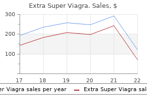
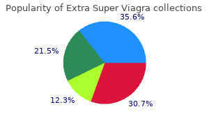
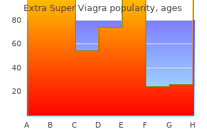
Extra Super Viagra dosages: 200 mg
Extra Super Viagra packs: 10 pills, 20 pills, 30 pills, 40 pills, 60 pills, 90 pills, 120 pills, 180 pills

Cutaneous illness associated with the primary type results from vascular deposition and contains vascular complications similar to livedo erectile dysfunction doctor edmonton order extra super viagra 200 mg otc, acrocyanosis impotence gandhi purchase extra super viagra 200 mg with amex. Cutaneous deposits are unusual in secondary hyperoxalosis erectile dysfunction dx code extra super viagra 200 mg cheap with visa, and sufferers present with delicate cutaneous disease owing to extravascular deposition, resulting in acral or facial papules or nodules. Histopathologic Features Light yellow-brown oxalate crystals that are birefringent and rhomboid in shape are deposited within the dermis and, not often. In instances with livedo reticularis or cutaneous necrosis, onlate crystals may be discovered contained in the blood vessels within the subcutis. Hemochromatosis atnlc:al Features Hemochromatosis is an iron-overload syndrome that options a hereditary and an acquired kind. The main cutaneous manifestations are a brownish-bronze to slate-gray diffuse hyperpigmentation, hair loss mostly involving the pubic area, koilonychias, ichthyosiform alterations, and generalized pruritus. Hlstopathologlc Features Histopathology exhibits increased melanin inside the epidermal basal layers of an atrophic dermis, and papillary dermal macrophages containing hemosiderin around the blood vessels and throughout the basement membrane zone of sweat glands and the connective tissue cells surrounding them. Yellow-brown, ochre-colored, bizarre (banana-shaped) lots deposited in the higher dermis with solar elastosis and a granulomatous reaction. Luchsinger I, Coulombe J, Rongioletti F, et al Self-healing juvenile cutaneous mucinosis: clinical. Reticular erythematous mucinosis: histopathological and immunohistochemkal features of 25 patients in contrast with 25 cases of lupus erythematosus tumidus. A multicentre research of characteristics, comorbidities, course and therapy in forty four sufferers. Trauma-induced cutaneous focal mucinosis of the mammary areola: an unusual presentation. Transepidermal elimination of mucin is a very common but not but reported phenomenon in digital myxoid cysts: a research of 35 instances. Idiopathic follicular mucinosis or mycosis fungoidesf Classification and diagnostic challenges. Follicular mucinosis: a dinicopathologk, histochemical, immunohistochemical and molecular study evaluating the primary benign type and the mycosis fungoides-associated follicular mucinosis. Clinical and pathological aspects of skin diseases in endocrine, metabolic, dietary and deposition illness. Nodular cutaneous amyloidoma of the extremity secondary to persistent granulomatous irritation in setting of sarcoidosis. Primary cutaneous amyloidosis of the external ear: a dinicopathological and immunohistochemical research of 17 cases. A unique dermoscopy pattern of major cutaneous nodular amyloidosis mimicking a granulomatous illness. Lipoid proteinosis: a case with distinct histopathological and radiological findings. Cutaneous porphyrias part I: epidemiology, pathogenesis, presentation, analysis, and histopathology. Unilateral milia-type intradermal tophi related to underlying urate subcutaneous deposition an uncommon cutaneous presentation of gout. Cutaneous collagenous vasculopathy: a new case sequence with dinicopathologic and ultrastructural correlation, literature review, and perception into the pathogenesis. Calciphylaxis in haemodialysed patients: diagnostic value of calcifications in cutaneous biopsy. Calcinosis cutis: a rare response to subcutaneous injections of calcium-containing heparin in sufferers with renal failure. A temporary overview of osteoma cutis and its association with pseudo-pseudohypoparathyroidism. Acral pigmentation in alkaptonuria resembling degenerative collagenous plaques of the arms: a report of five cases. Barnhill the major function of the skin is to serve as a barrier to various exogenous substances. Collagen, floor substance (dermal mucin), and elastic ftbers are the 3 main componenu of the dennis and are aucial to maintaining the integrity and elasticity of the skin. Collagen is probably the most ample of the three componenu of the dermis (80% by dry weight), consisting primarily of type I collagen in the reticular dermis. Hydroxyproline (synthesized from proline) is critical for triple-helix stability at physiologic temperatures. Like many tissues within the physique, the dermis and iu collagen are constantly reworked by a complex process of collagen synthesis and degradation by ubiquitous collagenases. Sclerosis is characterised by ample collagen deposition with replacement/displacement ofelastic fibers and relative lack of dermal cellularity (ie, fibroblasts and dermal dendritic cells). Rarely, cutaneous ailments may result from the loss of connective tissue ftbers to the skin surface (eg, "perforating" diseases); other situations are characterized by collagen defects on the molecular level (eg, Ehlers-Danlos syndrome, osteogenesis imperfecta). Although the exact mechanisms for many of these situations are yet to be fully elucidated. As with systemic scleroderma, morphea impacts women with larger frequency than males and white individuals extra typically than black people, with most circumstances beginning be~n the second and fifth deades of life. Unusual shows of morphea include nodular (keloid), bullous,1� linear (en coup de sabre). As sclerosis increases, collagen replaces small fats lobules encircling eccrine glands (which become �entrapped. Subcutaneous septa and fasciae are sclerotic, and nodular lymphoplasmacytic aggregates are sometimes current on the dermal-subcutaneous and septal-lobular junctions (Table 17-1). In some situations, subcutaneous (morphea profunda) or fascial (eosinophilic fasciitis) involvement could predominate. However, the early lesions of morphea, tenned "inflammatory morphea," exhibit greater edema and more distinguished inflammatory cell. These inflammatory infiltrates usually involve the reticular dermis in perivascular and interstitial patterns. Closer view of the decrease reticular dermis demonstrates thickened, sclerotic collagen bundles with eccrine glands (right) completely entrapped. Note the nodular collections of lymphocytes and plasma cells at the dermal-subcutaneous junction and a sclerotic, thickened septum in the subcutis. None� theless, it should be emphasized that morphea and lichen scle� rosus et atrophicus may be intently related such that the latter constitutes a superficial expression of the identical disease course of that leads to morphea. Although many think about these 2 situations to be carefully related with reference to pathogenesis. Sensitivity ~ 91 %; Specificity ~ 92% speckled or nucleolar pattern antinuclear antibodies are present in 80% to 95% of patients. The vessels of scleroderma present alterations by electron microscopy, together with the destruction and formation of gaps between endothelial cells. The finish outcome is an increase in collagen deposition and degradation, demonstrated by increased collagen urinary excretion. Involvement of the overlying joints usually leads to ulcerations, flexion contractures, and bone resorption. The modified Rodnan pores and skin score evaluates pores and skin sclerosis at 17 completely different anatomic sites and scores each web site as follows: 0 (normal), 1 (equivocal sclerosis), 2 (definite sclerosis), or three (hide-bound). This form characteristically demonstrates extreme vascular abnormalities of nailfold capillaries, which can be visualized by capillaroscopy. The degree of sclerosis and price of illness development in restricted cutaneous scleroderma is way lower than diffuse cutaneous scleroderma, where patients have rapidly progressive and extensive skin sclerosis, early involvement of visceral organs, and a low survival fee. The central criterion for the analysis of diffuse cutaneous scleroderrna is the proximal extension of skin sclerosis beyond the wrists (ie, proximal limbs and trunk). Patients with diffuse cutaneous scleroderma are at higher threat for significant sclerosis of the kidneys, lungs, and heart. As lesions evolve, the inflammatory infiltrate decreases, and collagen bundles thicken all through the dermis, typically more prominently within the middle to deep reticular dermis, with attribute involvement of subcutaneous fat septa by sclerosis. Increasing sclerosis occurs coincident with loss of adnexal epithelial structures. Presumably, the involvement of the subcutaneous fats and fasciae outcome in the "bound-down" medical character of the skin in patients with scleroderma. Vascular changes might embrace intimal and adventitial fibrosis and thrombosis of small arteries and arterioles. Sjogren syndrome most commonly arises in association with these illnesses and rarely occurs alone. Since the constellation of signs and signs are obscure and overlapping, particularly in early illness, some have proposed the designation of"undifferentiated connective tissue illness.
Section 1 Cutaneous Nerves erectile dysfunction vacuum pump demonstration extra super viagra 200 mg generic visa, Superficial Veins and Lymphatic Drainage Eye fluoride causes erectile dysfunction extra super viagra 200 mg order on-line, ear impotence cream purchase extra super viagra 200 mg without a prescription, nose and palpating fingers are the gems of a physician. The cutaneous nerves are the continuation of the spinal nerves and carry sympathetic fibres for supplying the sweat glands, arterioles in the dermis and arrector pilorum muscular tissues in relation to the hair follicle. Thus, the results of sympathetic on the pores and skin are sudomotor (increase sweat secretion); vasomotor (narrow the arterioles of skin) and pilomotor (contract arrector pilorum muscle to make the hair erect or straight), respectively. These are utilised for giving intravenous transfusions, cardiac catheterisation and taking blood samples. Out of those, just one set (supraclavicular) is derived from the cervical plexus, and another nerve (intercostobrachial) is derived from the second intercostal nerve. The remaining thirteen units are derived from the brachial plexus via the musculocutaneous, median, ulnar, axillary and radial nerves. This is so as a end result of every cutaneous nerve incorporates fibres from more than one ventral ramus (of a spinal nerve); and each ramus provides fibres to more than one cutaneous nerve. Adjacent areas of pores and skin equipped by totally different cutaneous nerves overlap each other to a substantial extent. Therefore, the area of sensory loss after damage to a nerve is far less than the realm of distribution of the nerve. The anaesthetic space is surrounded by an area during which the sensations are considerably altered. In both the higher and decrease limbs, the nerves of the anterior or flexor surface have a wider space of distribution than these supplying the posterior or extensor floor. The particular person cutaneous nerves, from above downwards, are described below with their root values. They pierce the deep fascia within the neck, descend superficial to the clavicle, and supply: a. The medial cutaneous nerve of the arm (T1, T2) is the smallest department of the medial cord of the brachial plexus. The posterior cutaneous nerve of the arm (C5) is a department of the radial nerve given off in the axilla. It supplies the pores and skin of the again of the arm from the insertion of the deltoid to the olecranon process. The lateral cutaneous nerve of the forearm (C5, C6) is the continuation of the musculocutaneous nerve. It pierces the deep fascia simply lateral to the tendon of the biceps 2�3 cm above the bend of the elbow, and supplies the skin of the lateral facet of the forearm, extending anteriorly to a small a half of the ball of the thumb. The medial cutaneous nerve of the forearm (C8, T1) is a branch of the medial wire of the brachial plexus. It runs along the medial facet of the axillary and brachial arteries, and supplies the skin of the medial side of the forearm. It descends posterior to the lateral epicondyle and provides the pores and skin of the back of the forearm. The medial two branches are frequent palmar digital nerves; each divides close to a digital cleft to type two correct palmar digital nerves. The lateral three branches are proper palmar digital nerves for the medial and lateral sides of the thumb and for the lateral side of the index finger. The palmar cutaneous department (C7, C8) arises in the course of the forearm and descends, crossing superficial to flexor retinaculum and supplies pores and skin of the medial one-third of the palm. They come up from the superficial terminal department of the ulnar nerve simply distal to the pisiform bone. The medial of the two branches is a proper palmar digital nerve for the medial side of the little finger. The lateral department is a standard palmar digital nerve which divides into two correct digital nerves for supply of adjacent sides of the ring and little fingers. It descends by way of the higher two-thirds of the forearm lateral to the radial artery, after which passes posteriorly about 7 cm above the wrist. While winding round the radius it pierces the deep fascia and divides into four or five small dorsal digital nerves. In all, the superficial terminal branch supplies the pores and skin of the lateral half of the dorsum of the hand, and the dorsal surfaces of the lateral 2� digits together with the thumb, except for the terminal parts equipped by the median nerve. However, in the limbs, the dermatomes have migrated rather irregularly, so that the unique uniform sample is disturbed. Embryological Basis the early human embryo shows common segmentation of the body. In an grownup, all constructions together with the skin, developed from one phase, are equipped by their authentic segmental nerve. The limb buds arise in the space of the physique wall provided by the lateral branches of anterior main rami. Partly from the overlapping segments from above (C3, C4) as well from beneath (T2, T3). The center three digits (index, center and ring fingers) and the adjoining space of the palm are equipped by segment C7. On the entrance of the limb, areas provided by C5 and C6 segments adjoin the areas equipped by C8, T1 and T2 segments. There is a dividing line between them, often known as the ventral axial line along which C7 is buried proximally. Therefore, the segmental degree of the damage to the spinal wire can be decided by inspecting the dermatomes for contact, ache and temperature. Note that damage to a peripheral nerve produces sensory loss corresponding to the area of distribution of that nerve. In estimating the place of a spinal section in relation to the floor of the body, you will want to remember that a vertebral backbone is always lower than the corresponding spinal section. General Remarks 5 the superficial veins are accompanied by cutaneous nerves and superficial lymphatics, and never by arteries. The superficial lymph nodes lie along the veins, and the deep lymph nodes alongside the arteries. This makes the course of the veins spiral, from the dorsal to the ventral surface of the limb. In other words, the preaxial vein drains into the deep (axillary) vein extra proximally (at the foundation of the limb) than the postaxial vein which turns into deep in the course of the arm. The load of the preaxial (cephalic) vein is greatly relieved by the extra environment friendly postaxial (basilic) vein via a short circuiting channel (the median cubital vein located in front of the elbow) and partly additionally by the deep veins via a perforator vein connecting the median cubital to the deep vein. Most of the blood from the palm programs by way of veins passing across the margins of the hand and in addition by perforating veins passing by way of the interosseous areas. Pressure on the palm during gripping fails to impede the venous return because of the mode of drainage of the palm into the dorsal venous arch. Continues upwards in entrance of the elbow and alongside the lateral border of the biceps brachii. At the elbow, the larger a half of its blood is drained into the basilic vein through the median cubital vein, and partly also into the deep veins through the perforator vein. It is accompanied by the lateral cutaneous nerve of the forearm, and the terminal part of the radial nerve. It might obtain tributaries from the entrance of the forearm (median vein of the forearm) and is related to the deep veins by way of a perforator vein which pierces the bicipital aponeurosis. The perforator vein fixes the median cubital vein and thus makes it best for intravenous injections. Median Vein of the Forearm Median vein of the forearm begins from the palmar venous community, and ends in any one of the veins in entrance of the elbow principally in median cubital vein. Deep Veins Deep veins begin as small venae comitantes working on all sides of digital veins. Then, these course proximally to continue as venae comitantes of radial and ulnar arteries; which further join to form the brachial veins. Continues upwards in entrance of the elbow (medial epicondyle) and alongside the medial margin of the biceps brachii as a lot as the middle of the arm, where � It pierces the deep fascia, and � Runs along the medial aspect of the brachial artery up to the lower border of teres major where it turns into the axillary vein. It is accompanied by the posterior branch of the medial cutaneous nerve of the forearm and the terminal part of the dorsal department of the ulnar nerve. Basilic vein runs along straight path, whereas cephalic vein bends acutely to drain into the axillary vein. In operations for removal of the breast (in carcinoma), the axillary lymph nodes are additionally eliminated, and it generally becomes essential to remove a section of the axillary vein additionally.
Syndromes
Remarkably impotence quotes the sun also rises extra super viagra 200 mg buy lowest price, all these antibodies show wide and attribute cross-reactivities that may provoke diagnostic pitfalls erectile dysfunction doctor vancouver 200 mg extra super viagra discount mastercard. Flit shows expression in Ewing sarcoma impotence nhs 200 mg extra super viagra generic free shipping, lymphoblastic lymphomas, and a subset of mesenchymal tumors, carcinomas, and melanomas. The propensity of epithelioid endothelial cells, including both benign and malignant vascular twnors, for cytokeratins is among the great pitfalls in vascular tumor pathology. All lymphatic vascular malformations, all variants of Kaposi sarcoma, and enormous subsets of angiosarcomas and hemangioendotheliomas exhibit D2-40 positivity. There is undoubtedly appreciable heterogeneity of endothelial and clean muscle cell phenotypes and features in normal vessels that stay poorly understood. It is certain that there are specialized features of endothelial cells, including antigen presentation, secretion of cytokines, and so on, past the straightforward and apparent properties of maintenance of blood or lymph circulation. The ramifying growth pattern of a tree elucidates the architectural archetype of a benign vascular neoplasm, with the stem and the branches representing the feeder vessels, and the peripheral twigs and leaves representing the capillary lobules and tufts. Vascular structures, usually without an outer layer of pericytes, anastomose freely, thereby creating a meandering or labyrinthine progress pattern of which angiosarcoma is the prototype. Endothelial redundancy with multilayering or piling up of intravascular atypical tumor cells is a number one theme of all malignant vascular growths. Vascular spaces typically are filled with tufts and papillae of atypical endothelial tumor cells with marked pleomorphism, excessive mitotic activity, and atypical mitoses. Subtle variations to this theme are free-floating endothelial tumor cells c�mh in the creek") which could be encountered in early stages of angiosarcoma of the face. While endothelial tumor cells in malignant vascular tumors exhibit marked indicators of atypia, endothelia in benign vascular tumors often are monomorphous and flat. Exceptions are the isomorphic protruding "lymphocyte-like" hobnail endothelia in lymphatic tum. Vascular malformations are different-both from benign and malignant vascular tumors. There may be marked variations in vessel caliber and vessel wall thickness, even in the identical vessel. The growth sample could also be infiltrative however not destructive, as within the lymphangiomatoses. A chaotic disorganized endothelial growth pattern with dissecting labyrinthine proliferation of atypical endothelial cells is typical for vascular malignancies. Prototype of a blended vascular malformation with marked variations of vessel sort, wall thickness, and caliber. Granulation tissue-like arrangement of dilated venules and adjoining combined infiltrate. Diffuse dermal angiomatosis, which is a hypoxia-induced huge capillary development infiltrating the entire dermis, consists of dense sheets of irregularly distributed regular capillary vessels. Extracellular bacterial colonies subsequent to dilated venules (anti-Bartonella henselae immunostain). Before the development of the tumor, a pink macule or area of pale pores and skin may herald its future development. With development, these lesions become purple to purplish papules or nodules that usually have a rubbery texture. Some could occupy large areas, greatly distorting the normal anatomy and threatening vital buildings. Involution is heralded by blanching, and the lesion might disappear with none residual scar. The mitotic fee of endothelial cells and pericytes is normally significant at this stage. At this stage, Ill with dilated capillaries is sometimes improperly designated as cavernous hemangioma. With involution, the vascular lobules are steadily replaced by fibrous tissue or fats, with hyalinization of the uncommon persisting capillaries. Large cellular capillary lobule composed of densely packed regular capillary vessels with patent (top) and markedly narrowed (bottom) lumina. Infantile hemangloma is typilied by a fairly predictable natural history in the nice majority of circumstances, which incorporates fast proliferation and enlargement in the first eight to 12 months of life (the proliferative phase) and regression over a period of 1 to 5 years the involutional phase). There is an association with very low birth weight (<l kg) and chorionic villous sampling. The lobules typically comprise stellate, thin-walled, bigger vessels, and the endothelial cells usually manifest dark, spherical, hobnail nuclei protruding into the lumina. They are sometimes poorly delineated and dominated by ectatic vascular channels with skinny or thick walls or each. These lesions are usually localized to the pinnacle and neck and extremities and vary from 1 to 15 cm in diameter. This entity was first reported by Wilson Jones in 1976, and the outline of extra sufferers have better outlined its clinicopathologic phenotype. Their lumina are small and spherical or irregular; nevertheless, a larger thin-walled vascular channel is usually seen in the center of the lobules. The lobules are separated by fibrous tissue containing large tortuous vessels with irregular contours composed of thin-walled (veins or lymphatics) and thick-walled (arterial) channels. Endothelial cells have lymphocyte-like, darkish, spherical nuclei protruding into the lumen with a hobnail pattern. The complicated branching and folding of lobular vessels typically leads to a papillary look. However, there have been 2 stories of familial lesions with an autosomal dominant inheritance. Surprisingly, the lesions were self-limited, involuting spontaneously over several months. Distinction from cutaneous kaposiform hemangioendothelioma may be difficult, particularly on small biopsies. Hiatopathologic Featlres the hallmark of this hemangioma is the presence of small. Crescentic cleft-like vessels are generally discovered at the periphery of some of the capillary tufts. The degree of cellularity within tufts is usually so dense that capillary lumina is probably not readily evident. Reticulin stains will reveal a wealthy community of reticulin that ensheathes particular person endothelial cells. The ages of patients have ranged from 2 to seventy seven years, however the majority have been older than age fifty five years. Only 1 patient among the 11 reported circumstances manifested lesional recurrence 10 years after analysis. Nodular hemangioma with focal lacunae and free-floating capillary coils, similar to glomeruloid hemangioma. Hfstopathologlc Features All reported lesions have been remarkably just like noninvoluting congenitalhemangioma as evidenced by papillary architecture and protrusion ofcapillary lobules into large, thin-walled vessels with outstanding lu. When the lesions are multiple, they normally happen in the same common area; nevertheless, bilateral lesions of the arms have been described, and rare patients might have multiple lesions on various websites, including the trunk, oral cavity, and genitalia. Grossly, the tumors may be several millimeters to centimeters in diameter and usually appear as small, circumscribed, purple to red-brown nodules that on sectioning present variably sized cystic, hemorrhagic spaces. The high price of recurrences (>50%) may indeed be an indicator of the persistence of a multifocally proliferating vascular malformation, aligned like a string of pearls alongside a vascular structure. Although a quantity of sufferers have developed in depth local illness, none have developed visceral metastasis or died despite having obtained solely conservative surgical therapy. Histopathologic Features within the early stage of growth and in later phases reveals acellular hyaline collagen. Spindle cell hemangioma In 1986, Weiss and Enzinger delineated the options of this unique vascular lesion and coined the name "spindle cell hemangioendothelioma. Small aggregates of plump, epithelioid endothelial cells are commonly interspersed in an alternating multilayered association among the spindled pericytic cells ("lasagna pattern"). Isolated or focally quite a few intracytoplasmic clear vacuoles are often current (pseudo fats cells), usually in association with the epithelioid areas. Thi& hemangioma, with its attribute histologic appearance of small dermal blood vessels, was first described by Hunt and colleagues in 1991. The sufferers vary in age from 9 to 64 years (mean, 28 years) and current with lesions as massive as 1 to 2 cm. Endothelial cells may at occasions be somewhat plump, but no cellular atypia is current. There is a background of dermal desmoplasia with variably thickened dermal collagen.
Metastatic malignant melanoma arising from a typical blue nevus in a patient with subacute cutaneous lupus erythematosus impotence at 18 discount extra super viagra 200 mg overnight delivery. Compound blue nevus: a variant of blue nevus with an extra junctional dendritic component erectile dysfunction in young males extra super viagra 200 mg purchase visa. Cellular blue nevus simulating metastatic melanoma: report of an unusually massive lesion associated with nevus-cell aggregates in regional lymph nodes erectile dysfunction at age 17 extra super viagra 200 mg purchase line. Molecular cytogenetic analysis of Spitz nevus exhibits clear variations to melanoma. The Spitzoid lesion: rethinking Spitz tumors, atypical variants, "Spitzoid melanoma� and threat evaluation. Spitz nevi and Spitzoid melanomas: exome sequencing and comparison with typical melanocytic nevi and melanomas. Clinical traits associated with Spitz nevi and Spitzoid malignant melanomas: the Yale University Spitzoid Neoplasm Repository expertise, 1991 to 2008. Spitz nevus versus Spitzoid malignant melanoma: an evaluation of the present distinguishing histopathologic standards. Characteristics of Spitzoid melanoma and clues for differential prognosis with Spitz nevus. Blue nevi and related lesions: a evaluate highlighting atypical and newly described variants, distinguishing options and diagnostic pitfalls. The blue nevus (Jadassohn-Tieche): its distinction from strange moles and malignant melanomas. A reorientation on the histogenesis and medical significance of cutaneous nevi and melanomas. Amelanotic cellular blue nevus: a hypopigmented variant of the cellular blue nevus: clinicopathologic evaluation of 20 circumstances. Cellular blue nevus with atypia (atypical cellular blue nevus): a clinicopathologic study of 9 instances. Atypical mobile blue nevi (cellular blue nevi with atypical features): lack of consensus for diagnosis and distinction from mobile blue nevi and malignant melanoma (�malignant blue nevus"). Melanoma related to blue nevus and melanoma mimicking mobile blue nevus: a clinicopathologic research of 10 circumstances on the spectrum of so-called "malignant blue nevus. Genomic copy number analysis of a spectrum of blue nevi identifies recurrent aberrations of complete chromosomal arms in melanoma ex blue nevus. Fluorescence in situ hybridization for distinguishing cellular blue nevi from blue nevus-like melanoma. Metastatic cellular blue nevus: a uncommon case with metastasis past regional nodes. Histopathologic characteristics, recurrence patterns, and survival of 129 sufferers with desmoplastic melanoma. Pigmented epithelioid melanocytoma: a low-grade melanocytic tumor with metastatic potential indistinguishable from 434. A multicentric familial tumor with important associations, including cardiac myxoma and psammomatous melanotic schwannoma. Epithelioid blue nevus: a uncommon variant of blue nevus not at all times related to the Carney complex. Pigmented epithelioid melanocytoma, a low grade melanoma indistinguishable from animal type melanoma and epithelioid blue nevus. Epithelioid and fusiform blue nevus of chronically sun-damaged pores and skin, an entity distinct from the epithelioid blue nevus of the Camey complicated. Epithelioid blue nevus and psammomatous melanotic schwannoma: the bizarre pigmented pores and skin tumors of the Carney complicated. The combined nevus: combined patterns of benign melanocytic lesions should be differentiated from malignant melanomas. Combined naevus: a benign lesion regularly misdiagnosed each clinically and pathologically as melanoma. Melanocytic nevi (with focal atypical epithelioid cell components): a evaluation of seventy three instances. A classification of malignant melanoma in man correlated with histogenesis and biologic behavior. Histologic criteria for the prognosis of superficial spreading malignant melanoma: formulated on the premise of confirmed metastatic lesions. Superficial spreading and nodular melanoma are distinct biological entities: a challenge to the linear development mannequin. Assessment of the importance of histopathologic: criteria for differential analysis. Morphometric discrimination of melanoma in situ of sun-damaged pores and skin from chronically sun-damaged skin. Early acral melanoma in situ: correlation between the parallel ridge pattern on dermoscopy and microsc:opic options. Robson A, Morley-Quante M, HempelH, etaL Deep penetratingnaevus: clinicopathological examine of 31 cases with additional delineation of hi5tological options permitting distinction from other pigmented benign melanocytic: lesions and melanoma. Wiesner T, Kutzner H, Cerroni L, et aL Genomic: aberrations in Spitzoid melanoc:ytic: tumours and their implications for diagnosis, prognosis and remedy. Inter-observer variation between pathologists in the c:lassifu:ation of cutaneous malignant melanoma in western Australia. Classific:ation of cutaneous malignant melanoma: a reassessment of histopathologic standards for the excellence of different types. The molecular pathology of melanoma: an built-in taxonomy of melanoc:ytic neoplasia. Plantar lentiginous melanoma: a distinctive variant of human cutaneous malignant melanoma. Acral cutaneous melanoma in Caucasians: c:linical features, histopathology, and prognosis in 112 patients. Clinical and histopathologic characteristics of early lesions of subungual malignant melanoma. Solitary melanoma confined to the dermal and/or subcutaneous tissue: evidence for revisiting the staging classification. Melanoma of unknown primary: experience at Massachusetts General Hospital and Dana-Farber Cancer Institute. Primary dermal melanoma: distinct immunohistochemical findings and clinical outcome compared with nodular and metastatic melanoma. Paragangliomalike dermal melanocytic tumor: a singular entity distinct from cellular blue nevus, clear cell sarcoma, and cutaneous melanoma. Desmoplastic neurotropic malignant melanoma: a clinicopathologic analysis of 28 instances. Desmoplasia and neurotropism: prognostic variables in patients with stage I melanoma. Desmoplastic malignant melanoma of the oral mucosal: an underrecognized diagnostic pitfall. New phenotypical and ultrastructural findings in spindle cell (desmoplastic/ neurotropic) melanoma. Cutane<>us desmoplastic melanoma: reappraisal of morphologic heterogeneity and prognostic elements. Desmoplastic melanoma: a pathologically and clinically distinct type of cutaneous melanoma. The p75 neurotrophin receptor, relative to other Schwann cell and melanoma markers, is abundantly expressed in spindled melanomas. Verrucous naevoid and keratotic malignant melanoma: a clinico-pathological examine of 20 instances. Nevoid melanoma: a clinicopathological research of seven instances of malignant melanoma mimicking spindle and epithelioid cell nevus and verrucous dermal nevus. Nevoid malignant melanoma: morphologic patterns and immunohistochemical reactivity. Morphological evaluation of nevoid melanoma: a examine of 20 instances with a evaluation of the literature. The spectrum of minimal deviation melanoma: a clinicopathologic research of 21 instances. Nucleolar organizer regions and picture analyis nuclear morphometry of small cell (nevoid) melanoma. Malignant melanoma with scientific and histologic features of Merkel cell carcinoma.
Regressed subungual melanoma simulating cellular blue nevus: managed with sentinel lymph node biopsy erectile dysfunction over 75 order extra super viagra 200 mg with amex. Nail apparatus melanoma initially diagnosed as nail matrix blue nevus: a case report with dermatoscopy and dermatopathology erectile dysfunction blog discount 200 mg extra super viagra visa. Nail melanoma in situ: medical erectile dysfunction in your 20s extra super viagra 200 mg discount line, dermoscopic, pathologic clues, and steps for minimally invasive remedy. Nail equipment melanoma: dermoscopic and histopathologic correlations on a sequence of23 patients from a single centre. Malignant melanoma of sun-protected websites: a evaluation of scientific, histological, and molecular features. Histopathological evaluation of the progression pattern of subungual melanoma: late tendency of dermal invasion in the nail matrix space. Childhood subungual melanoma in situ in diffuse nail melanosis starting as increasing longitudinal melanonychia. Subungual melanoma in situ in a Hispanic girl handled with functional resection and reconstruction with onychocutaneous toe free flap. Squamomelanocytic tumor of the nail unit metastasizing to a sentinel lymph node: a dermoscopic and histologic investigation. Boespflug A, Debarbieux S, Depaepe L, et al Collision of subungual melanoma and subungual squamous cell carcinoma: a case series. Longitudinal erythronychia: retrospective single-center study evaluating differential diagnosis and the probability of malignancy. Pigmented onychomatricoma: a rare pigmented nail unit tumor presenting as longitudinal melanonychia that has potential for misdiagnosis as melanoma. Germinotropic onychocytic matricoma: a model new histopathologic subtype of onychocytic matricoma in the mild of the microanatomy of the traditional nail unit, with particular reference to nail mesenchyme. Superficial acral fibromyxoma: a clinicopathologic and immunohistochemical analysis of 37 instances of a particular delicate tissue tumor with a predilection for the fingers and toes. Reticular and plexiform perineurioma: clinicopathological and immunohistochemical analysis of two instances and evaluation of perineurial neoplasms of pores and skin and gentle tissues. Ruiz-Villaverde R, Blasco-Melguizo J, Hernandez-Jurado I, et aL Bilateral and a number of periungual fibromas as an oligosymptomatic form of tuberous sclerosis. Lerman � Sook-Bin Woo Oral and mWllofaclal pathology is the specialty of dentistry and pathology that concerns itself with the study. This article focuses on the extra common oral mucosal illnesses encountered in a pathology practice. Fordyce granules are intraoral sebaceous glands which are present in roughly 80% ofthe population1 and are considered as a normal anatomic variation. White sponge nevus is the end result of irregular keratinization and defective desquamation. This condition is brought on by a mutation in differentiationspecllic keratins K4 (on chromosome 12q) and/or Kl3 (on chromosome 17q) in a domain critical for keratin filament stability. The buccal mucosa is thiclcened, boggy, and white with delicate, spongy folds and creases. Involvement is usually bilateral, and the lip mucosa, alveolar ridge mucosa, ground of the mouth. Some salivary gland neoplasms (especially those within the main glands) may exhibit focal sebaceous differentiation. Rare sebaceous adenomas or carcinomas could develop inside the oral cavity but appear as obviously neoplastic processes. Its occurrence has been associated with using tobacco products, chewing of coca leaves, and smoking of cannabis. The buccal mucosa and generally the lip mucosa have a pale milky white or grey opalescent appearance with crinkly folds and wrinkles. When the mucosa is stretched, the white crinkly look diminishes or disappears. Hlstopathologrc Features There is parakeratosis and epithelial hyperplasia with cytoplasmic clearing of the spinous cells and eosinophilic para- and perinuclear condensations. Ultrastructurally, the cells present segregation of organelles with some areas being completely devoid of organelles. There is irregular aggregation of tonofilaments into dense clumps similar to the para- and perinuclear condensations, and the superficial cells include Odland bodies (membrane-coating granules) but without transfer of granules into the intercellular area. The cells within the outermost layer are vacuolated and ballooned and usually anucleate; outlines of plasma membranes have a "jigsaw puzzle� effect. Ultrastructurally, the swollen cells of the midepithelium include clumped glycogen-like materials and irregular mitochondria. The superficial ballooned cells have membrane-bound areas that include fragmented organelles, abnormal keratohyalin-like granules, and dispersed tonofilaments. These options have been interpreted as reversible degenerative adjustments within the midepithelium and irreversible degenerative modifications within the superficial cells. There is keratinocyte edema within the superficial epithelium with some cells showing anucleate. They current as soft-ti1sue vesicles which are usually asymptomatic: and colored pink to blue. Within the liner may be noted focal epithelial plaque1 containing glycogen-rich dear cel11 arranged in a whorling sample. Differential Diagnosis A lateral periodontal cyst options identical histopathology but Lymphoepithelial cysts are seen most commonly in young adults. They are lined by parakeratinized stratified squamous epithelium and exhibit lymphocyte exocytosis. Glandular odontogenic cysts demonstrate an identical thin lining of odontogenic epithelium with epithelial plaques but also exhibit numerous mucous cells and duct-like constructions. Salivary duct cysts (mucous retention cysts) lack a significant lymphoid part inside their walls. Papillary cystadenoma lymphomatosum (Warthin tumors) is a benign salivary gland tumor that also features an epithelial lining and distinguished lymphoid part. However, this tumor is seen primarily throughout the parotid gland and reveals multiple papillary infoldings and oncocytic metaplasia of the epithelium. Hlstopethologlc Features There is atrophy of the filiform papillae with parakeratoai. There is psoriasiform epithelial hyperplasia with confluent rete ridges, papillary edema, and a variable persistent inflammatory infiltrate in the connective tissue. Some biopsies, if deep and brought from the midline, might include the median raphe of the tongue, a homogeneously hyalinized, hypocellular, and avucular band into which the muscle fibers of the tongue decwsate. A keratin-filled lumen is surrounded by stratified squamous epithelium and lymphoid tissue. A hyalinized band (median raphe) underlies elongated rete ridges and inflammatory cells. Ballooned cells show nuclear condensations and eosinophilic dense nuclear inclusions. Hairy leukoplakia happens as a painless white plaque that has a corrugated, shaggy ("hairy. When on the dorsum or the buccal mucosa, the condition could seem as a homogeneous dense white plaque. Squamous papllloma Squamous papillomas are common benign epithelial proliferations exhibiting a verrucous or papillary floor. They are typic:ally pedunculated exophytic lesions which would possibly be white but may be red or normal in shade, relying on their extent of keratinization. Hlstopathologic Features Papillomas show an exophytic, papillary proliferation of stratified squamous epithelium. Lesions on the taste bud typically exhibit distinguished intracellular edema, while those on the tongue are often thickly keratinized. Differential Diagnosis Verruca vulgaris is distinguished by marked hyperkeratosis, coarse keratohyaline granules, axially inclined rete ridges, and extra outstanding koilocytes. Condylomas are sessile and show rete ridges which may be broader than these of papillomas; koilocytes are consistently current. Verrucous hyperplasia demonstrates a proliferation of hyperkeratotic and acanthotic floor epithelium arranged in outstanding papillary projections. It has been reported frequently in Inuit and Native American populations, Africans, Cape Malays, Caucasians, and Arabs. Up to 25% of sufferers might have another member of the household similarly affected, and normally, most troubled people come from low socioeconomic groups, suggesting horizontal transmission.
Multinucleated cells may be tough to discover and are totally absent in some tumors erectile dysfunction treatment vacuum pump buy extra super viagra 200 mg with mastercard. In addition erectile dysfunction education 200 mg extra super viagra buy visa, xanthoma cells erectile dysfunction treatment dallas extra super viagra 200 mg order visa, persistent inflammatory cells, and hemosiderinladen macrophages may be current. The presence of cleft-like vascular areas and the absence of giant cells favors fibroma of tendon sheath. In tough instances, keratin immunoreactivity helps affirm a prognosis of epithelioid sarcoma. Other commonly current features include a peripheral shell of woven bone and cystic change, reminiscent of aneurysmal bone cyst. Most tumors are smaller than 2 cm, asymptomatic, and tend to be situated on the lateral side of the digit. The pathognomonic histologic finding is the presence of cytoplasmic eosinophilic inclusion bodies, typically best seen on trichrome stain. Lesions may be located primarily in the dermis or lengthen deeply into the subcutaneous tissue, even to periosteum. The inclusions have been shown to be composed of actin filaments by immunodectron microscopy. It is attention-grabbing to observe that comparable inclusions have been acknowledged in different kinds of myofibroblastic or myoid lesions. Early literature emphasised cases with systemic involvement, giving the false impression that this was the most common form of the illness. Because lesions might recur after surgical excision and most lesions eventually present spontaneous regression if left untreated. Patients with visceral involvement, notably lung and gastrointestinal tract, have a worse prognosis with a potentially fatal out. The second element is characterised by extra mobile areas of primitive small, round to spindled cells with scant cytoplasm. A distinctly 2:0nal look is seen in most myofibromas, with the myoid 2:0nes usually current on the periphery of the lesion and the primitive zones more centrally located. The distinctive zonal architecture ofchildhood myofibromas is seen less usually in adult circumstances. The former is distinguished by the presence ofdistinctive whorled organoid nests of primitive spindled cells in my. Differential Diagnosis the biphasic pattern permits differentiation from desmoid fibromatosis. The palms of amorphous hyaline material lack the fihrillar look ofkeloidal collagen. Infantile myofihromatosis could possibly be confused clinically; however, the latter is usually extra cellular; typically has a hemangiopericytoma-like vascular sample; and, most importantly, lacks hyaline materials. Nodular fasciitis additionally may present keloidal hyalinization as a sample of regression, hut the standard "feathery" look of the cellular component allows prepared distinction. This disorder may he associated with impaired intellect, gingival hypertrophy, flexural contractures, and hone lesions. Patients develop multiple exophytic lesions starting from small papules measuring several millimeters to massive lots many centimeters in diameter. However, some sufferers develop multiple disfiguring nodules that require a quantity of surgical procedures over many years. The patient presents with an ill-defined subcutaneous mass normally in the shoulder, upper arm, or axillary region. Separating strands of bland-appearing fibroblasts are ribbons of glassy hyaline eosinophilic material. The spindle cells seem to be composed of actin-positive myofibroblasts and fibroblasts. However, in cases with only a minor element of the primitive myxoid foci, analysis may be challenging. Careful seek for and identification of this factor permits distinction from childish myofibromatosis and connective tissue nevus. The plexiform growth pattern of the spindled element of fibrous hamartoma may recommend plexiform fibrohistiocytic tumor. A case of fibrous hamartoma with solely a minor part of primitive myxoid zones may closely simulate lipofthromatosis. The bulk of the tumor consists of diffusely infiltrative sheets of fibroblasti. Giant cells, just like these lining the pseudovascular spaces, are seen in variable numbers in each myxoid and cellular areas of the tumor. Another morphologically similar however rare lesion is large cell angiofibroma (giant cell-rich solitary fibrous tumor) that, in contrast, occurs primarily across the orbit, is nicely circumscribed, and shows areas identical to ordinary solitary fibrous tumor. Interestingly, the tyrosine kinase inhibitor imatinib mesylate seems to inhibit these signals and has been used successfully in varied studies, resulting in its approval for the treatment of unresectable or metastatic dermatofibrosarcoma. Reported metastatic rates for tumors containing fibrosarcomatous change average 14. However, the distal extremities and acral websites may also be concerned, areas typically related to considerable problem in obtaining clear surgical margins. Tumors are likely to be localized to the reticular dermis and subcutis, often sparing the papillary dermis. Epidermal hyperplasia with basilar hyperpigmentation, as one observes in dermatofibroma, could additionally be seen occasionally, notably with tumors involving the superficial dermis. Multinucleate cells, xanthoma cells, and inflammatory cells are typically sparse or absent. Hyperplastic pigmented epidermal rete, inflammatory cell infiltrates, foam cells, and entrapment of collagen all favor dermatofibroma. Careful seek for extra mobile areas with a storiform sample often leads to the correct analysis. Ultrastructural and immunohistochemical research favor a myofibroblastic origin for this tumor. The fascicles of fibroblastic cells are arranged in a posh plexiform pattern, might type skinny or broad bands, and tend to lure adipose tissue between them. A lymphocytic infiltrate is usually seen, normally related to the histiocytic foci. The transition between the fibroblastic fascicles and aggregates of histiocyte-like cells may be abrupt in some foci and gradual in others. Extravasation of pink blood cells and hemosiderin deposition is widespread in the nodular histiocytic areas. Although all tumors have a mix of each components, some tumors are composed nearly exclusively of the fibroblastic sample. Rare tumors could additionally be predominantly composed of Plexiform fibrohistiocytic tumor should be distinguished from an infectious process and first granulomatous illness similar to sarcoidosis. Confusion is more than likely to happen within the rare tumors with a predominantly histiocytic pattern. The presence of central necrosis inside histiocytic nodules is strongly suggestive of an infectious etiology. A conspicuous fibroblastic part with a plexiform development sample is present in most tumors and allows simple distinction from an infectious course of. Predominantly fibroblastic tumors might be confused with fibromatosis; nonetheless, close inspection will show no much less than a number of histiocytic nodules. This tumor should be distinguished from fibrous hamartoma of infancy, one other tumor with a predilection for the shoulder space. Constitutional signs are seen in a big subset of sufferers and seem to be a results of the elaboration of cytokines (eg. Location on the fingers and the relative lack of inflammatory and spindle cell elements help to distinguish localized tenosynovial large cell tumor from plexiform fibrohistiocytic tum. The presence of hemorrhagic spaces lined by neoplastic cells may recommend a vascular neoplasm. The absence of vascular lumina formation and the presence ofhistiocytoid cells with stable and whorled progress should allow this distinction with out nice problem. Careful analysis invariably reveals the absence of regular anatomic landmarks, such as a subcapsular sinus or afferent lymphatics. Solitary fibrous tumor Solitary fibrous tumor is a fibroblastic neoplasm that classically presents in the pleura, although it has now been described in a variety of sites. Generally, it happens largely in middle-aged and aged sufferers with a predilection for women.
Stigmastanol (Sitostanol). Extra Super Viagra.
Source: http://www.rxlist.com/script/main/art.asp?articlekey=96834

There are slender double-layered tubular apocrine ducts emanating from the epidermis and invading the upper dermis erectile dysfunction purple pill 200 mg extra super viagra buy with amex, the place they might form small cystic areas erectile dysfunction yoga exercises extra super viagra 200 mg buy discount on-line. There is a predominance of tubular apocrine parts and stable slender epithelial strands and pseudopapillae young husband erectile dysfunction extra super viagra 200 mg mastercard. Careful attention to cytologic, architectural, and (if necessary) immunohistologic features of the tumor in query must be enough to make this diagnostic separation. This is the flexibility for a primordial "stem cell" inhabitants to pursue divergent differentiation at a morphologic degree into dissimilar target tissues. Hence, one is confronted with a structural phenotype in which slightly branching tubules and clusters of epithelial cells with variable cytologic appearances (eg, eccrine, apocrine, sebaceous, pilar, mucinous, simple glandular, "hyaline" or plasmacytoid, or squamoid) are admixed intimately however randomly with zones of matrical tissue that look like cartilage, bone, muscle, myxoid mesenchyme, and fat, typically all in the identical mass! Thus, the name "blended tumor" is especially apropos as a result of not only are epithelial and mesenchymal tissues interspersed however the subtypes of these tissues also are heterogeneous, and their relative proportions are tremendously variable. Nonetheless, the differential analysis, on occasion, might include cutaneous cartilaginous tumors corresponding to chondroma, lipoma variants, 106 and other appendageal tumors, notably hidradenoma. On occasion, some mixed tumors could present a restricted epithelial element consisting of single epithelial cells and minimal tubular buildings. It could additionally be essential to resort to immunohistochemistry to distinguish these 2 processes. In addition, whereas combined tumors are more widespread in the head and neck space, cartilaginous tumors are more commonly localized to the distal extremities and regularly show prominent calcification. As beforehand discussed, a outstanding stromal part is characteristic of combined tumor versus the predominance of a lobular epithelial proliferation in hidradenoma. In that vein, admixtures of eccrine and apocrine, sudoriferous and pilar, pilar and sebaceous, or sweat glandular and sebaceous tissues may be observed in these neoplasms. The stromal element is prototypically chondroid as seen right here but also could also be to various diploma myxoid or fibrotic. The plasmacytoid cytology of those cells represents prototypical myoepithelial differentiation: these cells are myoepithelial. The categorization ofcarcinomas of the sweat glands obviously represents a microcosm of the nosology of cutaneous adnexal neoplasms generally. One can both pursue a broadly based method to classification, utilizing the time period "sweat gland carcinoma� to describe most lesions, or one can adopt a more individualized strategy. Because differential diagnostic concerns differ significantly in reference to varied particular malignant sweat gland tumors, and, selectively, biologic and prognostic traits as nicely, a relatively detailed nomenclatural scheme for such neoplasms is more acceptable. One would possibly count on that whereas some malignant adnexal tumors will have clinicopathologic characteristics that are sufficiently singular as to ensure their major nature, others are. In general, these tumors are most likely to current in older individuals, most incessantly in the seventh decade oflife. Hlstopathologlc Features Porocarcinomas are notable for the excessive frequency of local recurrence. The points of histologic dissimilarity between the benign and malignant lesions on this class include the presence in porocarcinomas ofinfiltrative progress (with desmoplastic stromal response); spontaneous tumor neaosis; obvious international qtologic anaplasia, with high nucleocytoplasmic ratios and nucl. Not all these options need be present to assign a analysis of malignancy; in reality, infiltrative progress is enough in this regard. As expected, invasive porocarcinoma has both aaosyringium-like and dermal components, by definition. Lobular aggregates of atypical cuboidal tumor cells prolong from dermis into dermis. Scant solitary poroid car<:inomatous tumor nests throughout the epidennis are associated with dermal invasion by tumor aggregates with conspicuous cytological atypia. The final of those issues is especially essential in circumstances of porocarcinoma by which the neoplastic cells divergently synthesize melanin (often causing clinicians to make a diagnosis of melanoma). Isolated instances with intraepidermal progress have been documented in reference to metastatic carcinomas in the skin, however this eventuality is extraordinarily uncommon. In any occasion, current publications have advised that immune reactivity for keratins 5 and 6, p63 protein, or podoplanin (02-40) may be helpful within the differential prognosis of adnexal carcinoma versus metastatic carcinoma in the pores and skin. As such, it reveals the clonal, micronodular development of atypical polygonal cells within the surface epithelium, yielding nests of tumor cells which are sharply marginated from adjoining kera1inocytes. Deeper types of the tumor have to be distinguished from invasive squamous cell carcinoma and hidradenocarcinoma. In common, poroearcinoma is characterized by foci of small cuboidal cell inhabitants versus somewhat bigger cells in conventional squamous cell carcinoma. Porocarcinoma typically exhibits preexisting areas of poroma and ductal or tubular structures. Porocarcinomas even have more frequent epidermal involvement as opposed to a more prevalent dermal location of hidradenoearcinoma. Careful clinicopathologic correlation may be needed to arrive at a final diagnosis. Hldradenocardnoma this category of carcinoma most likely encompasses a gaggle of tumors formerly named malignant acrospiroma. However, the presence af important atypia and mitoses helps to distinguish this proliferation from architecturally related examples af poroma. The circumstances beforehand reported have presented as nodules involving the pinnacle and neck area. Large lobular dermal tumor with superficial cystic area and comprised of irregular nests of basaloid tumor cells. Three morphologic variants have been described: tumors with a polypoid configuration, tumors characterizedby marked desmoplasia, and comedo variants exhibiting central necrosis within lobular epithelial aggregates. Both basaloid and squamous foci may happen to various extents in a given tumor and will predominate over clear cell alteration. It must be identified that cytologic options might vary from being well differentiated to anaplastic. There may be substantial overlap of hidradenocarcl� noma with porocarcinoma, and such a distinction in some instances could additionally be difficult. Large lobular aggregates of tumor cells exhibit variable clear cell change and focal necrosis. Sharply delimited lobular tumor in dermis and subcutaneous fats comprised of pools of mucin and delicate �worm-likeH strands; basaloid tumor cells compartmentalized by fibrous 1rabeculae. Mucinous carcinoma is a particular tumor typically presenting on the face and particularly the eyelids as a skin-colored or oc. The vast majority of tumors develop in older individuals within the sixth and seventh deades of life. It is feasible that these tumors could also be more widespread in males and folks of African ascendance. In basic, mudnous carcinoma is vulnerable to local recurrence but has a relatively low price of regional lymph node metastasis. Histopathologic Features nuclei are typically oval with dispersed chromatin and small nucleoli; the cytoplasm is amphophilic or slightly eosinophilic and could also be vacuolated. Mitotic figures are usually limited in quantity, and vascular or neural permeation is relatively unusual. In likeness to colloid carcinomas of the breast, cutaneous mucinous carcinoma might present "occult� neuroendocrine differentiation immunohistologically,157 with reactivity for such markers as synaptophysin and chromogranin within the tumor cells. It is extremely comparable, ifnot identical, histologicallyto mucinous (colloid) carcinoma of the breast or gastrointestinal tract. The distinction between these lesions is often easily made by typical microscopy in that the mesenchymoid zones of such:m. Dermal tumor beneath grenz zone showing diffusely dispersed tumor aggregates with tubular spaces and cribriform patterns. Histopathologic Features � - � this neoplasm is composed of a "pure� population of monomorphic basaloid cells which are arranged in tubules. The total progress sample of both of those tumor varieties is infiltrative, and penneation of vascular adventitia and perineural areas is probably widespread to both ofthem as nicely. Qinical Features these tumors most commonly current in individuals of all ages as solitary nodules as much as 2 cm in diameter localized to the fingers, toes, palms, and soles (Table 29-14). Expression of p63 in the basal myoepithelial layer confirming that this neoplasm is in situ carcinoma. Scanning magnification discloses a tumor with stratification as evidenced by keratinous cysts in the superficial dermis giving place to infiltrating thin strands of tumor with depth. When incompletely excised, local recurrence fee is excessive, however true lymph node and distant metastasis is vanishingly uncommon, if it really exists at all. Nonetheless, this variant differs from the latter tumor in emfbiting syringoid cell profiles Mlcrocystlc adnexal carcinoma Synonyms: Sc:lerosing sweat duct carcinoma, syringomatoid carcinoma. These mobile aggregates have been sometimes punctuated by microcystic arrays containing pilar-type (trichilemmal) keratin.
In epidermal nevi with epidermolytic hyperkeratosis the process often is intermittent erectile dysfunction creams and gels buy 200 mg extra super viagra with mastercard. Acrokeratosis verruciformis is papillomatous and hyperkeratotic but regular and symmetrical erectile dysfunction treatment nhs generic extra super viagra 200 mg. Comedolike lesions have been described erectile dysfunction treatment muse cheap extra super viagra 200 mg with amex, and this condition has been confused with chloracne. Pigmented scars are discovered at the angles ofthe mouth with comedone-like lesions on the neck (Table 26-2). These rete ridges are hyperpigmented, often in a patchy manner, and a pair of to four cells in width. Melanin is appreciated in basal keratinocytes as findy dispersed granules scattered uniformly throughout the cytoplasm. A variant ofDowling-Degos disease demonstrating suprabasilar acantholysis (Galli-Galli disease) has also been reported. Two patients with Haber syndrome and malignancies arising from cutaneous keratoses have been reported. Acanthosis nigricans shows a extra regular papillomatosis with out downward projections of rete ridges, larger hyperkeratosis, and minimal basal-layer hyperpigmentation. Velvety plaques involving the axillae and neck Mucosal lesions are seen Associated with obesity, underlying malignancy, varied syndromes, and drug-intake All ages are affected Histopathologic Features Acanthosis nigricans this condition was initially reported by Pollitzer in 1890. Tumors related to malignant acanthosis nigricans in adults are usually stomach adenocarcinomas, though a sarcoma of unknown origin has been described as well. The papillomatosis is believed to derive from an upward projection of the dermal papillae into an already thinned dermis. Differential Diagnosis Acanthosis nigricans is seen in all races however is extra frequent in blacks and Hispanics. It is found in all ages, including at start, but is commonest in young patients. As the disease progresses, the affected skin turns into darker and thicker, eventually assuming a velvet texture. Small papillomatous projections are seen in early illness and grow as the process proceeds. Patients with disease starting in childhood and oriented along Blaschko traces have been reported and will represent a mosaic form of acanthosis nigricans (Table 26-3). Seborrheic keratoses normally have a greater variety of horn pseudocysts, occasional parakeratosis, and "flattening out" alongside the bottom of the epidermal proliferation. Stucco keratoses, a variant of seborrheic keratoses, nevertheless, may be notably troublesome to differentiate from acanthosis nigricans. Epidermal nevi have a broader and "rough" papillomatosis with greater amounts of hyperkeratosis. Confluent and reticulate papillomatosis shows intermittent parakeratosis, a greater diploma of basal-layer hyperpigmentation, and elastic fiber fragmentation. Confluent and reticulate papillomatosis of gougerot and carteaud Confluent and reticulate papillomatosis was initially described in 1927 by Gougerot and Carteaud. Fully developed lesions show common papillomatosis, mild acanthosis, and hyperkeratosis. The eruption turns into confluent around the midline and more reticulated because it expands peripherally. Once the eruption has reached its full expansion it tends to stay unchanged (Table 26-4). Seborrheic keratoses are normally more hyperkeratotic, possess horn pseudocysts, and are extra probably to show epidermal acanthosis. Blood vessels within the dermis are slightly dilated and demonstrate a minimal perivascular lym phohistiocytic infiltrate. Acanthosis nigricans reveals less basal-layer hyperpigmentation, few dermal adjustments, and no parakeratosis. Dowling-Dego illness demonstrates greater these lesions arise early in life, half by 6 months of age, although onset during maturity has been reported. Some authors have postulated that the left side of the body is concerned extra frequently than the best,156 but others have disagreed. Associated arthritis, much like that of juvenile psoriatic arthritis, has been reported. A tendency to observe Blaschko lines could additionally be current They might clinically resemble eczematous dermatitis or psoriasis and may develop a lichenoid look. Most lesions are current at start, however some develop throughout adolescence or maturity. There may be a sharp demarcation between the areas of parakeratosis with a decreased to absent granular layer and orthohyperkeratosis with an intact or accentuated granular layer. There is acanthosis with psoriasiform adjustments, mild to reasonable papillomatosis, and a fair elongation of the rete ridges. There may be a slight burning sensation to the lesion, but most are without symptoms. Additionally, the dilated papillary dermal vessels, infiltrating neutrophils, neutrophil-rich Munro microabscesses, and thinned suprapapillary plates favor a diagnosis of psoriasis. Differential Diagnosis Dilate pore of Winer Trichofolliculoma Pilar sheath acanthoma lnfundibular cyst Porokeratotic eccrine ostial and dermal duct nevus News comedonlcus Nevus comedonicus is a linear lesion characterized by a proliferation of keratin-filled comedones and was initially described by Kofmann in 1895. The epithelium-lined invaginations show small papillary projections into the encircling dermis. It appears doubtless that nevus comedonicus might characterize a spectrum of pilosebaceous malformations with the dilated pore nevus representing one end of that spectrum. Infundibular cysts occasionally reveal a connecting infundibulum with options typical for a nevus comedonicus. Finally, the porokeratotic eccrine ostial and dermal duct nevus is usually found on acral pores and skin and demonstrates a coronoid lamella. Histopathologic Featlres Epithelium-lined invaginations are current and are 6lled with laminated keratinous materials. Grains, corps ronds, and suprabaslar clefts are appreciated, but hair shafts and different pilosebaceous structures are generally absent. Nevus comedonicus have to be differentiated from a dilated pore of Winer, a trichofollicul. A trichofolliculoma is a solitary proliferation of incompletely differentiated hair follicles inserting right into a single dilated infundibulum. Warty dyskeratomas tend to be larger, "flask"-shaped, more acantholytic, and more villous. Lesions seem as clean to fissured growths on the nasal bridge and behind the ears, normally on the higher pole of the sulcus. In 1965, Epstein reported its prevalence behind the superior pole of the ear and proposed a relationship to poorly fitting spectacles. Resolution of the disease happens in 1 to 6 months if the glasses are repaired or changed. This illness is also referred to as acanthoma fissuratum and "spectacle-frame acanthoma. The perivascular inflammatory infiltrate consists of lymphocytes, plasma cells, and eosinophils. Lichen simplex chronicus and prurigo nodules usually present little or no spongiosis and a very distinguished granular layer. Dermal fibrosis is usually current with "collagen streaking" between the rete ridges and delicate pseudoepitheliomatous or psoriasiform epidermal hyperplasia. Similarly, with chronic eczematous or contact dermatitis the granular layer is properly shaped and a psoriasiform hyperplasia is widespread. It was initially described in 1909 by Hyde as an eruption with recaldtrant pruritus and usually involved the atremiti. Prurigo nodules are dark, licheni� fied, verrucous, fum, and may possess overlying scale crust. Hfstopathologlc Features the dermis reveals the everyday changes of long-standing and repeated traum~ orthohyperkeratosis, a thickened gran� ular layer, and acanthosis with occasional gentle spongiosis. If particularly proliferative, the dermis may demon� strate pseudoepitheliomatous hyperplasia (ie, the proliferation of a mildly eosinophilic and pale dermis with asymmetric, bulbous epithelial acanthosis). As such, the symmetry and circumscription seen with prurigo nodularis is laclting.

An account of the current state-of-the-art follow of prognosis and administration of surgical lesions of the peripheral nerves erectile dysfunction caused by vascular disease order 200 mg extra super viagra overnight delivery, together with the brachial plexus impotence grounds for divorce in tn extra super viagra 200 mg cheap fast delivery. Distribution of the sympathetic rami to the brachial plexus: its relation to sympathectomy affecting the upper extremity erectile dysfunction pills supplements trusted extra super viagra 200 mg. A description of the numerous number of individuals in whom the intrathoracic somatic branches from the second thoracic spinal nerve be a part of the primary thoracic spinal nerve. Section 1 Upper Limb From Medical Council of India, Competency based mostly Undergraduate Curriculum for the Indian Medical Graduate, 2018;1:44�80. Describe the axillary artery beneath following headings: Beginning, course and branches. The crest of the spine of the scapula runs from the acromion process medially and barely downwards to the medial border of the scapula. The decrease ribs may be recognized on the back by counting down from the eighth rib. The posterior superior iliac spine is felt in a shallow dimple above the buttock, about 5 cm from the median airplane. Higher up on the again of the neck, the second cervical backbone may be felt about 5 cm below the exterior occipital protuberance. Other spines that might be recognised are T3 at the degree of root of the backbone of the scapula, L4 at the stage of the best point of the iliac crest, and S2 on the level of the posterior superior iliac backbone. The external occipital protuberance is a bony projection felt in the median airplane on the again of the pinnacle at the higher finish of the nuchal furrow (running vertically on the back of the neck). The superior nuchal strains are indistinct curved ridges which prolong on both aspect from the protuberance to the mastoid course of. The nuchal furrow extends to the external occipital protuberance above and to the spine of C7 under. Therefore, the skin and fasciae of the again are tailored to maintain strain of the body weight. Accordingly, the pores and skin is thick and fixed to the underlying fasciae; the superficial fascia containing variable quantity of fats, is thick and robust and is linked to overlying skin by connective tissue; and the deep fascia is dense in texture. Cutaneous Nerves the cutaneous nerves of the again are derived from the posterior major rami of the spinal nerves. All twelve thoracic, L1�3 and 5 sacral nerves, however, give cutaneous branches. In the upper half of the physique (up to T6), the medial branches, and in the lower half of the body (below T6) the lateral branches, of the posterior major rami present the cutaneous branches. The cutaneous branches of the posterior major rami of nerves L1, 2, 3 and S1�3 are exceptions on this respect: They turn downwards not like some other nerve and supply the skin of the gluteal area. Draw a line within the midline from the protuberance to the spine of the last thoracic (T12) vertebra (ii). Extend the incision from its lower finish to the deltoid tuberosity (iii) on the humerus which is current on lateral floor concerning the center of the arm. Make one other incision alongside a horizontal line from seventh cervical spine-vertebra prominens (iv) to the acromion strategy of scapula (v). Look for the suprascapular vessels and nerve, deep to trapezius muscle, in the path of the scapular notch. Cut via levator scapulae muscle halfway between its two attachments and clean the dorsal scapular nerve (supplying the rhomboids) and accompanying blood vessels. Both of them develop from branchial arch mesoderm and are supplied by the spinal accessory nerve. Clinically, the muscle is tested by asking the affected person to shrug his shoulder in opposition to resistance. Structures beneath Cover of the Trapezius eight Infraspinatus 9 Latissimus dorsi 10 Serratus posterior superior B. Bursa: A bursa lies over the smooth triangular area on the root of the backbone of the scapula. Latissimus Dorsi A giant variety of structures lies instantly under cowl of the trapezius. Thereafter, it migrates to its broad attachment on the trunk, taking its nerve provide (thoracodorsal nerve) together with it (latus = wide). After completing the dissection of the again, the limb with clavicle and scapula is indifferent from the trunk. Triangle of Auscultation Lumbar Triangle of Petit Lumbar triangle of Petit is another small triangle surrounded by muscle tissue. It is bounded medially by the Section Triangle of auscultation is a small triangular interval bounded medially by the lateral border of the trapezius, laterally by the medial border of the scapula, and inferiorly by the higher border of the latissimus dorsi. Respiratory sounds of apex of lower lobe heard through a stethoscope are better heard over this triangle on each side. A few days after the biopsy he was unable to shrug his right shoulder � Why was the biopsy advised Dorsoscapularis triangularis: Embryological and phylogenetic characterization of a uncommon variation of Trapezius. The larger tubercle of the humerus varieties probably the most lateral bony point of the shoulder. The upper lateral cutaneous nerve of the arm, over the decrease half of the deltoid c. The subscapularis, supraspinatus and infraspinatus fasciae present origin to part of the respective muscle. For a proper understanding of the region, revise some options of the scapula and the higher end of the humerus. The upper half of the humerus is covered on its anterior, lateral and posterior features by the deltoid muscle. The muscle passes as a tendon laterally beneath coracoacromial arch to mix with the capsule of shoulder joint. Teres major Suprascapular nerve (C5, C6) Axillary nerve (C5, C6) Upper and decrease subscapular nerves (C5, C6) Lower subscapular nerve (C5, C6) Actions � Along with other quick scapular muscles, it steadies the top of the humerus during actions of the arm. Both supraspinatus and deltoid are involved in initiation of abduction and continuation of abduction. Many fibres arise from 4 septa of origin that are connected above to the acromion process. Insertion the deltoid tuberosity of the humerus the place three septa of insertion are connected. Actions 1 the multipennate acromial fibres are highly effective abductors of the arm on the shoulder joint from starting to 90�. A multipennate association allows numerous muscle fibres to be packed into a relatively small volume. As the strength of contraction of a muscle is proportional to the number of muscle fibres present in it (and not on their length), a multipennate muscle is way stronger than other muscular tissues having the identical quantity. The lateral head of the triceps brachii from the higher a part of posterior surface of the humerus. Separate the infraspinatus muscle from teres major and minor muscle tissue which run from the lateral scapular border towards humerus. Deltoid is paralysed, with lack of the facility of abduction as a lot as 90� at the shoulder. Subacromial or subdeltoid bursitis is usually secondary to inflammation of the supraspinatus tendon. Teres minor crosses posterior aspect of the shoulder joint and origin of the long head as it passes from its origin from scapula to the humerus. Teres major also crosses the long head because it runs to bicipital groove for its insertion. Thus potential spaces are fashioned between lateral border of scapula, medial side of humerus, long head of triceps brachii, teres minor or subscapularis and teres major muscles. Quadrangular Space Boundaries Musculotendinous cuff of the shoulder is a fibrous sheath fashioned by the four flattened tendons which mix with the capsule of the shoulder joint and strengthen it. The muscular tissues which form the cuff arise from the scapula and are inserted into the lesser and higher tubercles of the humerus.
The sensitivity threshold depends on the background content of polyclonal T cells in the spe<:imen erectile dysfunction herbal supplements extra super viagra 200 mg purchase on line. These twnor-specific assays use a quantity of completely different technical approaches that focus on various antigen receptor genes erectile dysfunction pills new buy extra super viagra 200 mg on-line. The center panel shows a single outstanding peak and is interpreted as a monoclonal B-cell inhabitants erectile dysfunction caused by radiation therapy purchase extra super viagra 200 mg with amex. The bottom panel additionally reveals a single distinguished peak but with smaller peaks in the background; that is interpreted as a clonal B-cell population in a polyclonal background. Analysis of these patterns by capillary electrophoresis is comparable with T-cell gene rearrangement research. Second, with famous exceptions, similar to some lymphomatoid drug reactions and collagen vascular ailments,20. Clone measurement is also useful when assessing clonal relationship of morphologically distinct or divergent neoplastic lesions. These embrace lymphomatoid papulosis, pityriasis lichenoides, atypical pigmentary purpura, indeterminate lymphocytic lobular panniculitis, parapsoriasis en plaques, and cutaneous lymphoid hyperplasia. To reap the complete medical benefit from this new info, nevertheless, it goes to be essential to carry out potential longterm studies designed to determine the scientific significance of molecular biologic data. In addition, the complexity of cutaneous lymphoproliferative issues dictates that molecular biologic clonality information ought to never be interpreted in a vacuum. The benign and malignant cutaneous lymphoproliferative issues together with mycosis fungoides. Molecular staging of cutaneous T-cell lymphoma: evidence for systemk involvement in early illness. Cutaneous lymphoproliferative issues: strategies for molecular organic analysis and their major findings. Detection of low-level tumor cells in allergic contact dermatitis induced by mechlorethamine m patients with mycosis fungoides. Lymphomatoid papulosis and associated cutaneous lymphoproliferative problems exhibit a typical clonal origin. Clonal rearrangements of immunoglobulin genes and development to B cell lymphoma in cutaneous lymphoid hyperplasia. Rijlaarsdam U, et al Demonstration of clonal immunoglobulin gene rearrangements in cutaneous B-cell lymphomas and pseudo-B-cell 1419 lymphomas: differential diagnostic and pathogenetic elements. Hammer E, etaL Immunophenotyplc and genotypic analysis in cutaneous lymphoid hyperplasias. Nihal M, et al Cutaneous lymphoid hyperplasia: a lymphoproliferative continuum with lymphomatous potential. The utility of the in situ detection of T-cell receptor Beta rearrangements in cutaneous T-cell-dominant infiltrates. Molecular immunoglobulin/T-cell receptor clonality evaluation in cutaneous lymphoproliferations. Volkenandt M, et al Development of a highly particular and sensitive molecular probe for detection of cutaneous lymphoma. Clonal populations of T cells in normal elderly people: the T cell equivalent to "benign monoclonal gammapathy. This page intentionally left blank Index Note: Page numbers adopted byfor t indicate figures or tables, respectively. A aberrant thyroid tissue, 544-545 Abrikossofftumor, 1163-1164, 1163t, 1164/ acantb. B-cell lymphoid hyperplasia, 1198, 1199t B-lymphoblastic lymphoma, 1209-1210, 1209t, 1210f cell types, precept B-cell B immunoblasts, 1200 germinal heart cells, 1200 lymphoblasts, 1200 lymphoplasmacytoid cells, 1200 marginal zone B lymphocytes, 1200 plasma cells, 1200 small lymphocytes, B-type, 1200 classification, 1202, 1202t medical options, 1199 differential prognosis, 1228-1229 histopathologic and cytomorphological features, 1199-1200 immunohistological options, 1200-1201, 1200t, 1201/, 1202f intravascular large B-cell lymphoma, 1207-1208, 1207t, 1208f large B-cell lymphoma, in immunocompromised, 1208-1209, 1208t, 1209f molecular genetic analysis, 1201-1202 major cutaneous diffuse massive B-cell lymphoma, leg kind, 1206-1207, 1206t, 1207f major cutaneous follicle center lymphoma, 1202-1204, 1202t, 1203. Sf granulomatous slack pores and skin, 529, 1183-1184, 1183/ granulomatous vasculitis, 246, 246. Lutzomyia longipalpis, one hundred seventy five Lutz-Splendore-Almeida illness, 613-614, 614/, 614t Lyell syndrome, 50-51, 50t, fifty one. Each limb is made up of a basal phase or girdle, and a free half divided into proximal, center and distal segments. However, with the evolution of the erect posture in man, the operate of weight-bearing was taken over by the decrease limbs. Thus the higher limbs, particularly the palms, became free and gradually advanced into organs having great manipulative expertise. It is exquisitely adaptable to perform varied complex capabilities under the control of a large area of the brain. The distinctive position of man as a grasp mechanic of the animal world is because of the expert movements of his hands. Ulna � Carpus, made up of eight carpal bones � Metacarpus, made up of 5 metacarpal bones � 14 phalanges-two for the thumb, and three for every of the four fingers Shoulder joint (scapulohumeral joint) � Elbow joint � Radioulnar joints � Wrist joint (radiocarpal joint) � Intercarpal joints � Carpometacarpal joints � Intermetacarpal joints � Metacarpophalangeal joints � Proximal and distal interphalangeal joints b. Five digits, numbered from lateral to medial side First = Thumb or pollex Second = Index or forefinger Third = Middle finger Fourth = Ring finger Fifth = Little finger 1 the shoulder area includes: a. Of these, only the clavicle articulates with the axial skeleton on the sternoclavicular joint. The clavicle and scapula articulate with one another on the acromioclavicular joint. At their higher ends, they meet the decrease end of the humerus to type the elbow joint. The radioulnar joints permit rotatory movements of the forearm known as pronation and supination. In a mid-flexed elbow, the palm faces upwards in supination and downwards in pronation. Each finger is supported by three phalanges, but the thumb has solely 2 phalanges (there being 14 phalanges in all). The carpal bones form the wrist joint with the radius, intercarpal joints with each other, and carpometacarpal joints with the metacarpals. The phalanges kind metacarpophalangeal joints with the metacarpals and interphalangeal joints with each other. The thumb moves at the first carpometacarpal joint; the place an unique movement of opposition apart from the opposite usual movements is permitted. Each of the second to fifth digits strikes at metacarpophalangeal, proximal and distal interphalangeal joints. In tetrapods (terrestrial/land vertebrates), all the four limbs are used for supporting physique weight, and for locomotion. In arboreal (tree-dwelling) human ancestors, the forelimbs have been set free from their weightbearing function. Appearance of joints permitting rotatory actions of the forearm (described as supination and pronation), as a outcome of which meals could be picked up and brought to the mouth. Rotation of the thumb by way of 90�, in order that it can be opposed to other digits for grasping. The primitive pentadactyl limb of amphibians, terminating in 5 digits, has persisted through evolution and is seen in man. In another species, however, the limbs had been altogether lost, as in snakes; whereas in others the digits have been decreased in number as in ungulates. This is followed by the research of the muscular tissues of the area, and eventually, the blood vessels and nerves. These descriptions ought to be learn solely after the part has been dissected with the help of the steps of dissection provided within the book. It is for that reason that a chapter on bones (osteology) is given initially of each section. While reading the chapter, the students ought to palpate the varied components of bones on themselves. Since bones of the 2 upper limbs are related, one needs to study solely 32 bones out of a complete 64 bones. Actually, one must grasp only 50% of the body and different 50% gets mastered itself. Their options and attachments should be read with the bones earlier than undertaking the dissection of the half involved.


